Magnetic Resonance Imaging

Magnetic resonance imaging (MRI) is the gold standard imaging modality for soft tissues. It produces detail cross-sectional images of soft tissue and bone anatomy, including muscles, tendons, ligaments, brain and organs, without the use of ionizing radiation. In addition to orthopedic imaging, MRI is also used for heart, brain and breast. MRI uses gadolinium contrast in many exams to highlight tissues and blood vessels, which enhances images and offers better diagnostic quality. It can also be used in conjunction with PET scans. How does MRI work? MR creates images by using powerful magnets to polarize hydrogen atoms in water (the body is made of of more than 80% water) so they face in one direction. A radiofrequency pulse is then used to ping these atoms, causing them to wobble, or resonate. The MRI coils detect this and computers can assemble images from the signals. Basic MRI scans will focus on the resonance of fat and water in two different sequences, which highlight and contrast different features in the anatomy.

Thumbnail

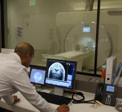
MRI effective for pediatric appendicitis, even when read by adult-focused radiologists

Ultrasound (US) and CT are typically viewed as the most appropriate imaging modalities for diagnosing pediatric appendicitis, but what about MRI?

Thumbnail

10.5T MRI scanners tested on humans as imaging equipment keeps getting bigger

After four years of testing on animals and making key updates, the University of Minnesota’s Center for Magnetic Resonance Research in Minneapolis was finally ready to test its massive 10.5T MRI scanner on a human. And once the scan was complete, it turned out it was worth the wait.

Thumbnail

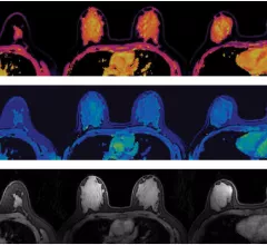
New technique brings MR fingerprinting to breast imaging

A new breast imaging technique uses MR fingerprinting for the volumetric quantification of T1 and T2 relaxation times for breast tissue, according to new research published in Radiology.

Thumbnail

FDA approves 7T MRI scanner at USC for clinical use

The 7T MRI scanner at the Keck School of Medicine of USC in Los Angeles has received FDA approval for clinical use.

Thumbnail

Installation of new MRI equipment leads to dozens of damaged iPhones, Apple Watches

It’s common knowledge that helium can make someone’s voice sound like a chipmunk, but the gas may be capable of another unexpected trick as well—doing serious damage to Apple products.

Thumbnail

MRI scans help researchers understand the biology of autism

With the help of MRI, San Diego State University researchers found the amygdala of children on the autism spectrum disorder (ASD) had weaker connections with some regions of the brain compared to “typically developing” children within the same age group.

MRI shows potential to correct slow gastric emptying, gastroparesis

Researchers have used MRI to show the impact of sending electrical pulses to the vagus nerve in an attempt to correct gastrointestinal problems and provide “more precise treatment” that pharmaceutical therapies and diet have not achieved.

Thumbnail

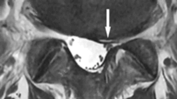
AI detects lumbar vertebrae in MRI images with 98.6% accuracy

A deep learning algorithm can automatically detect lumbar vertebrae in MRI images, according to findings published in the Journal of Digital Imaging. This, the authors noted, has potential to improve clinician efficiency.

Around the web

The nuclear imaging isotope shortage of molybdenum-99 may be over now that the sidelined reactor is restarting. ASNC's president says PET and new SPECT technologies helped cardiac imaging labs better weather the storm.

CMS has more than doubled the CCTA payment rate from $175 to $357.13. The move, expected to have a significant impact on the utilization of cardiac CT, received immediate praise from imaging specialists.

The all-in-one Omni Legend PET/CT scanner is now being manufactured in a new production facility in Waukesha, Wisconsin.