Magnetic Resonance Imaging

Magnetic resonance imaging (MRI) is the gold standard imaging modality for soft tissues. It produces detail cross-sectional images of soft tissue and bone anatomy, including muscles, tendons, ligaments, brain and organs, without the use of ionizing radiation. In addition to orthopedic imaging, MRI is also used for heart, brain and breast. MRI uses gadolinium contrast in many exams to highlight tissues and blood vessels, which enhances images and offers better diagnostic quality. It can also be used in conjunction with PET scans. How does MRI work? MR creates images by using powerful magnets to polarize hydrogen atoms in water (the body is made of of more than 80% water) so they face in one direction. A radiofrequency pulse is then used to ping these atoms, causing them to wobble, or resonate. The MRI coils detect this and computers can assemble images from the signals. Basic MRI scans will focus on the resonance of fat and water in two different sequences, which highlight and contrast different features in the anatomy.

Thumbnail

Brain fMRI can help mend broken hearts

Some break-ups are just so traumatic, a person can’t even see the point of getting out of bed. But one musician looked to neuroscience and fMRI to help stop a heavy heart from keeping her down.

Thumbnail

Australian government allots $125M for MRI but opponents call it 'window dressing'

Australia’s federal government is allocating approximately $125 million ($175 million Australian) for MRI. Prime Minister Scott Morrison and Health Minister Greg Hunt both back the funding and believe it will result in improved diagnosis and treatment.

ASRT shares new white paper on MR safety

The American Society of Radiologic Technologists (ASRT) has published a new white paper, Radiologic Technologist Best Practices for MR Safety, designed to help MR technologists provide the safest patient care possible.

Thumbnail

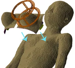
Could this research help prove cutting-edge MRI techniques are safe for patients?

Researchers have simulated how more than 20 different breast tissue ratios respond to heat emitted from MRIs at higher field strengths than those currently in hospitals, according to findings in Magnetic Resonance in Medicine.

Thumbnail

Kansas City hospital offers ‘abbreviated MRI’ to screen for breast cancer

Abbreviated MRI—a new breast imaging modality at University of Kansas Health System—makes screening quicker and less painful, according to a report by local NBC affiliate KSHB-TV.

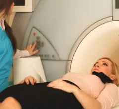
Physicist develops VR app to reduce children's fear, anxiety before MRI

Jonathan Ashmore is an MRI physicist with National Health Service, the United Kingdom’s public healthcare system. He doesn’t interact with patients too often, but with an office next door to radiology department, he heard the distress children experienced before, during and after an MRI exam.

Thumbnail

Breast MRI proves helpful with inconclusive digital mammography, DBT results

Radiologists can feel confident turning to breast MRI in instances when digital mammography (DM) and digital breast tomosynthesis (DBT) results are inconclusive, according to new research published in the American Journal of Roentgenology.

Thumbnail

Biannual MRI outperforms annual mammography for high-risk breast cancer patients

Biannual MRI is more effective than annual mammography in detecting early breast cancer in young women who have a high genetic risk of the disease, according to new research published in Clinical Cancer Research.

Around the web

The nuclear imaging isotope shortage of molybdenum-99 may be over now that the sidelined reactor is restarting. ASNC's president says PET and new SPECT technologies helped cardiac imaging labs better weather the storm.

CMS has more than doubled the CCTA payment rate from $175 to $357.13. The move, expected to have a significant impact on the utilization of cardiac CT, received immediate praise from imaging specialists.

The all-in-one Omni Legend PET/CT scanner is now being manufactured in a new production facility in Waukesha, Wisconsin.