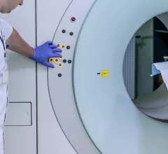
Magnetic Resonance Imaging

Magnetic resonance imaging (MRI) is the gold standard imaging modality for soft tissues. It produces detail cross-sectional images of soft tissue and bone anatomy, including muscles, tendons, ligaments, brain and organs, without the use of ionizing radiation. In addition to orthopedic imaging, MRI is also used for heart, brain and breast. MRI uses gadolinium contrast in many exams to highlight tissues and blood vessels, which enhances images and offers better diagnostic quality. It can also be used in conjunction with PET scans. How does MRI work? MR creates images by using powerful magnets to polarize hydrogen atoms in water (the body is made of of more than 80% water) so they face in one direction. A radiofrequency pulse is then used to ping these atoms, causing them to wobble, or resonate. The MRI coils detect this and computers can assemble images from the signals. Basic MRI scans will focus on the resonance of fat and water in two different sequences, which highlight and contrast different features in the anatomy.

Thumbnail

Australian hospital opens $21M MRI suite to support complex neurosurgery

Australia’s Sir Charles Gairdner Hospital has opened the doors on a $21-million MRI renovation after more than a year of construction, government officials announced this week.

Thumbnail

Safety update: What we know about performing MR exams on patients with implantable cardiac devices

Recent research has shown MRIs are safe for patients with implantable cardiac devices such as pacemakers and cardioverter defibrillators. However, according to a recent analysis published by Radiology, there’s still a lot of information for radiologists, cardiologists and electrophysiologists to know about this important topic

Thumbnail

High-strength MRI could release toxic mercury from silver dental fillings

High-strength MRI may release mercury, a known toxin, from amalgam fillings in teeth, according to a new study published in Radiology.

Thumbnail

University of Minnesota invests $2M to cut MRI wait times

Around $2 million has been funneled into the University of Minnesota’s latest healthcare project: a third MRI machine that’s expected to cut waiting times for patients who typically wait up to three weeks for a scan.

Thumbnail

Patient sent home because he was ‘too large’ for MRI machine sues hospital for $7M

An Oregon patient who was sent home after he failed to fit in an MRI machine is suing a local hospital for $7 million, citing medical negligence as the reason he was left with permanent paraplegia, the Portland Business Journal reported this week.

Thumbnail

New York patient discharges gun in MRI room, charged with reckless endangerment

A Long Island, New York, man was arrested last month after a handgun he possessed fired and injured him while in an MRI room, according to a June 20 report from the LI Herald.

Thumbnail

Radiology nurse fired after neglecting patient in MRI machine

A radiology nurse at Tartu University Hospital in Tartu, Estonia, was fired after a patient undergoing an MR exam after knee surgery was left inside the machine unattended for more than an hour.

Thumbnail

Long Island patient charged with reckless endangerment after gun discharges in MRI exam room

An MRI patient in Long Island, New York, was arrested last month after a firearm in his pocket discharged in the exam room, resulting in injuries to both of his legs, the LI Herald has reported.

Around the web

The nuclear imaging isotope shortage of molybdenum-99 may be over now that the sidelined reactor is restarting. ASNC's president says PET and new SPECT technologies helped cardiac imaging labs better weather the storm.

CMS has more than doubled the CCTA payment rate from $175 to $357.13. The move, expected to have a significant impact on the utilization of cardiac CT, received immediate praise from imaging specialists.

The all-in-one Omni Legend PET/CT scanner is now being manufactured in a new production facility in Waukesha, Wisconsin.