Magnetic Resonance Imaging

Magnetic resonance imaging (MRI) is the gold standard imaging modality for soft tissues. It produces detail cross-sectional images of soft tissue and bone anatomy, including muscles, tendons, ligaments, brain and organs, without the use of ionizing radiation. In addition to orthopedic imaging, MRI is also used for heart, brain and breast. MRI uses gadolinium contrast in many exams to highlight tissues and blood vessels, which enhances images and offers better diagnostic quality. It can also be used in conjunction with PET scans. How does MRI work? MR creates images by using powerful magnets to polarize hydrogen atoms in water (the body is made of of more than 80% water) so they face in one direction. A radiofrequency pulse is then used to ping these atoms, causing them to wobble, or resonate. The MRI coils detect this and computers can assemble images from the signals. Basic MRI scans will focus on the resonance of fat and water in two different sequences, which highlight and contrast different features in the anatomy.

Thumbnail

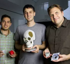
Novel 3D-printing technique generates full anatomical models from MRI, CT scans

A 3D-printing technique originated at Harvard University allows clinicians to produce highly detailed models of human anatomy in less than an hour—for a fraction of the cost and labor needed for a lower quality product, researchers reported in 3D Printing and Additive Manufacturing this month.

Amplified MRI captures brain movement in conjunction with heartbeat

Novel technology known as phase-base amplified MRI is the first to capture the brain’s movement in conjunction with a heartbeat, opening the door for earlier detection of brain disorders and abnormalities, researchers from Stanford University and the University of Auckland announced this week.

Thumbnail

PET/MRI with full head scan catches more brain abnormalities than standard protocol

It might not be the majority of hospitals’ standard routine, but including a patient’s head during routine body imaging with FDG PET/MRI could detect far more brain abnormalities than traditional practice, according to research published in the American Journal of Roentgenology.

Thumbnail

MRI, PET reveals how extreme athlete ‘The Iceman’ withstands sub-zero weather

Wayne State University radiologists have unlocked at least part of the puzzle behind extreme athlete Wim “The Iceman” Hof’s uncanny ability to withstand freezing temperatures—and it’s shed light on the degree of control the brain can have over the human body.

Lawsuit claims Detroit attorney had undisclosed financial ties to MRI center

A lawsuit filed by State Farm Insurance claims Detroit attorney Mike Morse had an undisclosed financial relationship with Berkley, Michigan-based Horizon Imaging, an MRI provider that accepted referrals from Morse's law firm.

Thumbnail

MRI could help transgender teens struggling with identity

The structure and activity in transgender teens’ brains more closely resemble their desired gender than their biological sex, according to research presented at the European Society of Endocrinology’s annual symposium in Barcelona.

Brain MRI could ID metastases in breast cancer patients, but guidelines don’t recommend it

Though not the guideline-recommended route for treatment, select breast cancer patients could benefit greatly from MRI screenings designed to identify brain metastases, according to a research letter penned this month by Dana-Farber/Brigham and Women’s Cancer Center experts and published in JAMA Oncology.

Thumbnail

Conventional MRI shows promise for measuring lung function in asthmatics

Conventional MRI has a new use in the clinical setting, Canadian researchers wrote in Radiology this month: measuring lung ventilation abnormalities in free-breathing patients.

Around the web

The nuclear imaging isotope shortage of molybdenum-99 may be over now that the sidelined reactor is restarting. ASNC's president says PET and new SPECT technologies helped cardiac imaging labs better weather the storm.

CMS has more than doubled the CCTA payment rate from $175 to $357.13. The move, expected to have a significant impact on the utilization of cardiac CT, received immediate praise from imaging specialists.

The all-in-one Omni Legend PET/CT scanner is now being manufactured in a new production facility in Waukesha, Wisconsin.