Magnetic Resonance Imaging

Magnetic resonance imaging (MRI) is the gold standard imaging modality for soft tissues. It produces detail cross-sectional images of soft tissue and bone anatomy, including muscles, tendons, ligaments, brain and organs, without the use of ionizing radiation. In addition to orthopedic imaging, MRI is also used for heart, brain and breast. MRI uses gadolinium contrast in many exams to highlight tissues and blood vessels, which enhances images and offers better diagnostic quality. It can also be used in conjunction with PET scans. How does MRI work? MR creates images by using powerful magnets to polarize hydrogen atoms in water (the body is made of of more than 80% water) so they face in one direction. A radiofrequency pulse is then used to ping these atoms, causing them to wobble, or resonate. The MRI coils detect this and computers can assemble images from the signals. Basic MRI scans will focus on the resonance of fat and water in two different sequences, which highlight and contrast different features in the anatomy.

AI artificial intelligence stethoscope doctor

After seeing 17 different doctors, boy with rare condition receives diagnosis from ChatGPT

The program landed on tethered cord syndrome, which a neurosurgeon later confirmed as the correct diagnosis. 

Thumbnail

Mayo Clinic, GE HealthCare launch strategic collaboration seeking to innovate in imaging and theranostics

Both will work in tandem to produce new research and products, aspiring to “transform the experience of patients and clinicians in the practice of radiology.” 

Thumbnail

Radiologists must be aware of side effects from new Alzheimer’s treatments, study warns

“As the use of monoclonal antibodies becomes more widespread, close collaboration between neurologists and radiologists is needed," one expert advised. 

Thumbnail

Accelerated MRI sequence reduces scan times by one-third while maintaining image quality

This change in clinical practice could increase scanner throughput, leading to decreased costs, patient discomfort and motion artifacts, experts noted. 

Medicare money payment physician

Optimizing imaging sites of service could boost margins, amass thousands more in collections

An organization could add an additional $87,532 on one MRI machine or over $260,000 via CT each year through strategic site selection, experts estimated.

Thumbnail

Introducing MRI into clinical practice helps reduce the rate of unnecessary prostate biopsies

“We argue that the results could be replicated elsewhere if guidelines are followed and the urologist and MRI radiologist are dedicated,” researchers noted. 

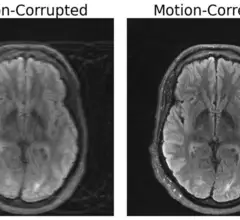
Motion-Corrected MRI Scan

MRI results affected by movement? MIT researchers have an AI-powered solution

An MRI's sensitivity to movement during scans often leads to image artifacts, risking misdiagnoses and redundant scans. But a new deep learning model may be able to compensate.

Fujifilm Echelon Synergy MRI

FDA clears new AI-powered MRI system from Fujifilm

The Echelon Synergy MRI machine utilizes proprietary deep learning reconstruction technology to enhance sharpness and acquire images faster. 

Around the web

The nuclear imaging isotope shortage of molybdenum-99 may be over now that the sidelined reactor is restarting. ASNC's president says PET and new SPECT technologies helped cardiac imaging labs better weather the storm.

CMS has more than doubled the CCTA payment rate from $175 to $357.13. The move, expected to have a significant impact on the utilization of cardiac CT, received immediate praise from imaging specialists.

The all-in-one Omni Legend PET/CT scanner is now being manufactured in a new production facility in Waukesha, Wisconsin.