Chinese team develops MRI-compatible robot to facilitate neurosurgery
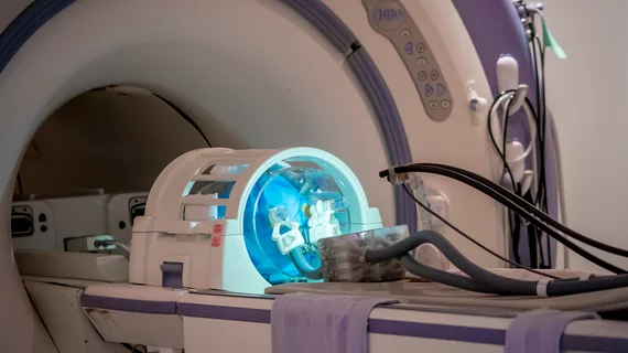
A team of Hong Kong scientists led by Kwok Ka-wai, PhD, have developed the world’s first intraoperative MRI-guided robot for bilateral stereotactic neurosurgery, opening new doors for less invasive, safer and more accurate treatment of conditions like Parkinson’s disease.
Stereotactic surgery has long been used to treat neuropsychiatric disorders, Ka-wai and colleagues at the University of Hong Kong said in a statement. Deep brain stimulation in particular is able to target specific sites of surgical interest with high specificity, allowing for the delivery of electrical signals to deep brain targets, but the procedure still suffers from surgical complications.
“This surgery is tremendously demanding on accuracy, by targeting only the tiny nucleus structures and not damaging the surrounding critical tissue,” the researchers said. “Without the intraoperative updates of a surgical ‘roadmap,’ the brain may shift after the skull is opened and inevitably lowers the targeting accuracy.”
Conventional deep brain stimulation is performed while a patient is under local anesthesia, so surgeons have to rely on verbal and physical interactions with that patient to ensure electrode placement is going smoothly.
Since illnesses like Parkinson’s are so widespread—it’s projected to affect more than 8.7 people worldwide by 2030, Ka-wai et al. said—“any improvement to this surgery would benefit a large population.”
The team devised its compact MRI-guided robot to facilitate less invasive stereotactic surgeries while a patient is under general anesthesia. With the robot, surgeons are able to more accurately control and evaluate the stereotactic manipulation bilaterally to the left and right brain targets in real time.
Robots usually contain some kind of electromagnetic motor, Ka-wai and colleagues said, which prevents them from coming too close to MRI machines. The researchers circumvented that by developing an MR-compatible tele-operated system driven by liquid, which still produces quality images during robot operation.
The research is also applicable to a host of other procedures, the researchers said, since its compact design fits into any standard MRI head coil.
“The success of this project would represent a major step toward safer, more accurate and effective brain surgery,” Ka-wai et al. wrote. “It is believed that all these components can be applied to other interventions requiring MRI guidance, like cardiac catheterization, prostate or breast biopsy.”
The team said it plans to conduct further studies on the subject. The authors' current research, which recently won the Best Conference Paper Award at the IEEE International Conference on Robotics and Automation, is published in IEEE Robotics and Automation Letters.

