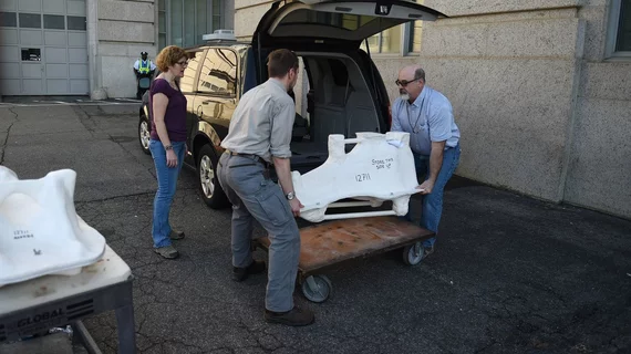
150-pound dinosaur skull gets CT scan for upcoming museum exhibition
It’s not every day that imaging equipment is used to scan a 68-million-year-old dinosaur skull, but that’s exactly what happened at George Washington University in Washington, D.C.
The 150-pound fossil, which belonged to an adult edmontosaurus, was transported in two separate pieces so that specialists could perform a CT scan, according to a report from the Washington Post. The scans are part of an upcoming exhibition at the Smithsonian National Museum of Natural History.
“The scanned skull will be featured in the digital display intended to explain how the dinosaur ate,” the article explained. “The hospital delivered the standard medical imaging files to the museum, which will use them to create a 3D version. The digital exhibit will showcase three dinosaur skulls and show how their bones, muscles, ligaments and tendons worked together.”
The skull was first collected back in 1931, the Washington Post reported, and is still in good condition.
Click below for the full story.

