MRI scans reveal 'big step' for progressive MS research
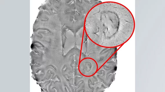
Chronic active lesions visible on brain MRI scans, identifiable by their “darkened outer rims,” are associated with multiple sclerosis, according to new findings published in JAMA Neurology.
“We found that it is possible to use brain scans to detect which patients are highly susceptible to the more aggressive forms of multiple sclerosis (MS),” senior author Daniel S. Reich, MD, PhD, of the NIH’s National Institute of Neurological Disorders and Stroke in Bethesda, Maryland, said in a prepared statement. “The more chronic active lesions a patient has the greater the chances they will experience this type of MS. We hope these results will help test the effectiveness of new therapies for this form of MS and reduce the suffering patients experience.”
Back in 2013, the researchers found that 7T MRI scanners could ID chronic active lesions, opening up the opportunity to take their work into new directions.
“Figuring out how to spot chronic active lesions was a big step and we could not have done it without the high-powered MRI scanner provided by the NIH,” first author Martina Absinta, MD, PhD, also from the NIH, said in the same statement. “It allowed us to then explore how MS lesions evolved and whether they played a role in progressive MS.”
The researchers scanned the brains of 192 MS patients, noting that 56% had at least one chronic active lesion. Of those patients, 44% had “rimless” lesions, 34% had one to three lesions and 22% had at least four lesions. The scans were then compared to exams the patients received when they first enrolled at the NIH’s Clinical Center. Patients with at least four of these lesions were 1.6 times more likely to be diagnosed with progressive MS than patients without the lesions. Those patients with at least four lesions also had less white matter and a smaller basal ganglia.
Patients who had undergone brain imaging once every year for 10 straight years, or longer, were also analyzed. When the rimless lesions shrunk in size, the authors found, the lesions with rims either grew in size or stayed the same size and were damaged.
“Our results support the idea that chronic active lesions are very damaging to the brain,” Reich said in the statement. “We need to attack these lesions as early as possible.”

