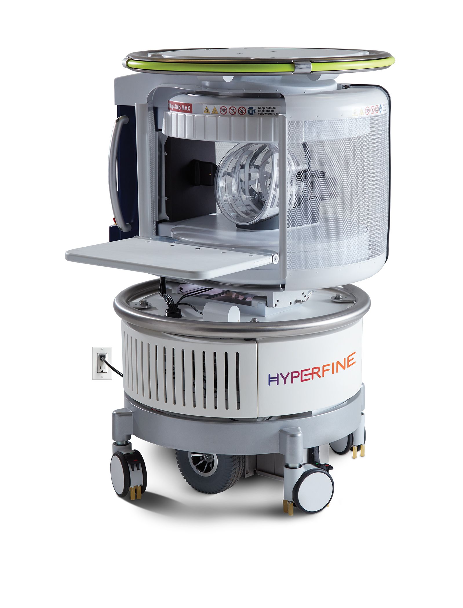
Built for value-based care: How the world’s first portable MR scanner is democratizing imaging
Connecticut-based Hyperfine made waves earlier this year when it announced the arrival of what it calls the “world’s first” portable MRI scanner. Which brings about interesting questions: How does it work? Is it replacing traditional technology? And what has COVID-19 meant for mobile imaging?

Chief Medical Officer
Hyperfine
Radiology Business recently sat down—over Zoom, of course—with radiologist Khan Siddiqui, MD, Hyperfine’s chief medical officer. He and colleagues plan to demonstrate the novel modality at RSNA’s annual meeting later this month.
Radiology Business: What is Swoop?
Siddiqui: Swoop is the world’s first FDA-cleared portable MR imaging system, which makes patient-centric imaging possible by bringing the scanner to the bedside, rather than the patient going to the scanner. It really started with our Founder Jonathan Rothberg’s vision to solve problems that we passionately care about in radiology: How do we actually build something that addresses a scenario that our loved ones have faced, and create a social impact at the same time? The big vision and mission is to democratize healthcare by making MR imaging accessible to everyone around the world. The whole idea started with: How do you get a scanner to the patient’s bedside? And the first thing that comes to mind is, well, it needs to go through a door. So that defines your size the device needs to be, right? And then suddenly that starts creating restraints that you are trying to solve. Jonathan put together a brilliant team in 2014 to address all of these challenging problems.
RB: How might this new bedside technology transform clinical workflows?
Siddiqui: Our goal really is to expand the reach of the radiology department out into the hospital and the community. Today on a daily basis, patients who are really sick, where it is too dangerous to move them, just don’t get imaged. Often the decision is made clinically—in the intensive care units, the emergency room, pediatrics, neuro-critical care, or neonatal ICU—where it’s just too difficult or too risky to move a patient anywhere. And a lot of decisions are subsequently made without enough data. The goal of the scanner is to enrich those decision-making processes.
These are just the hospital scenarios. The most common call to a nursing home is because somebody had slurred speech, dizziness or something of that nature, and an ambulance shows up and takes them to a hospital. Then you find out that the patient was dehydrated and now you have tallied $10,000 in costs for ambulance transport and a day of admission. Whereas with Swoop, you can do the scan right there and then and completely reduce the costs to the system. We literally are designed for value-based care.
RB: How does a low-field system like this fit within the conventional MRI world?

Siddiqui: If you look at the history of low-field imaging, that’s how scanners started. The early versions were all low field in the 1980s. And then GE commissioned the first 1.5 tesla scanner in the mid ’80s, and after that, everybody jumped ship to high field. But if you look at the improvements in image quality from that GE 1.5T scanner to now, it’s day and night. It’s all because of improvements in reconstruction technology, electronics, and the understanding of the physics behind it. And a lot of that research that was happening with 1.5 tesla and above never got tried on a low-field strength. Then about five or six years ago, there was a resurgence of interest, with improvement in electronics, deep and machine learning, artificial intelligence and the cloud. Suddenly, there was a lot more computing power available to extract signal and create images from techniques that were just not available in the ’80s. That’s what started this regeneration and now lower field strengths systems are a hot topic.
It solves a lot of problems. No. 1, you don’t need liquid helium or a ton of power to cool the magnet. And then because they are low-field strength, you have less requirements for gradient and RF amplifiers. That reduces power requirements even further. Our scanner is literally powered the same way as a coffee maker; you can just plug it into any power outlet. And with improvements in electronics and GPUs for processing, we can take the entire rack of servers that the typical scanner uses for processing and put them right underneath the chassis. Usually scanners have a separate computer rack, but we can compress all of that stuff into the electronics underneath.
RB: What are some of the implications of portable imaging and its impact on the larger hospital ecosystem?
Siddiqui: It’s a very simple unit to clean and disinfect, with very flat surfaces. Just following the instructions provided using sanitary wipes, in a few minutes you’ll have the whole device clean from an infection control point of view. MR scanners typically start at $1 million, and ours is $50,000. That is magnitudes less. Swoop is accessible and it is designed with very simple training. You don’t need to have an MR degree. You can have a radiology tech who does portable x-rays run the scanner, too. We’ve designed this in such a way that it can be controlled wirelessly through either a tablet or even remotely from a laptop. So, the tech can sit in the MR suite and control 10 different scanners at the same time. We’ve made it so simple that it’s like an iTunes playlist. You select why you want to do the scan, hit play, and that’s it. A nurse can do it, a physician can run it, anyone can. We’ve made it really, really simple and easy to use.
RB: What about reimbursement for this work?
Siddiqui: We are focused initially toward inpatient scenarios, so most billings are going to be DRG (diagnosis-related group) based. And we are working with some of the payers to define what the right reimbursement for this is going to be over time. So today, the sites are using the typical MRI billing codes. But I think the true value comes when there is dedicated reimbursement for this modality, which is different from what a full traditional scanner would do. Those are active discussions going on right now.
RB: What kind of traction are you getting in the market? How are customers responding to the technology?
Siddiqui: To be honest with you, I just got off of a call and there was an argument going on over who would get it first for a hospital site [laughs]. I get comments like, “We need two of them here, three of them there,” and it literally becomes: “How fast can you get it to us?” What I’ve noticed is it’s the people who are in the clinical workflows of those patients who immediately get the value, whether they’re in neurosurgery, neurology or obviously radiology.
RB: What else would you like to share about this solution?
Siddiqui: Our scanner is similar to what a portable or conventional x-ray does. It’s just increasing the access to it. Think of a trauma scenario: You immediately do a portable x-ray and once the patient is stable, you take them down and do the entire body all over again, and there is immediate value for that quick scan, so you can generate clinical value right away and see what is happening. Also, we are complementary to the existing MR, not conflicting with it. You just become part of the entire continuum of care for those scenarios. You can use the scanner as a monitor for a patient who has some sort of hemorrhage or edema. If you want to see if it’s improving or not, you can have a scanner in the patient’s room and every few hours, keep scanning just to see how it is improving and whether the hemorrhage is resolving or not.
RB: Has the coronavirus pandemic changed the conversation at all around Swoop?
Siddiqui: COVID-19 has proven to be a powerful scenario for this technology. There were patients who came in with shortness of breath, hyperventilated and then just stopped responding or improving. How do you figure out what is going on? Do you keep treating them aggressively? They’re unconscious, and we ended up finding strokes and bleeds and other things on scanners that would not be possible before. As you know, 70% of patients who are in the ICU for COVID end up having some neurological findings. So, it becomes very difficult to do that imaging.
And with this COVID, one of the big changes that’s happened is the greater acceptance of portable imaging, with much more of it being done in the outpatient setting. In 2020, so much has changed in healthcare, and one of the biggest trends is greater acceptance of portable imaging and being able to do imaging at the patient’s bedside.
To sign up for one of their RSNA events, or to learn more, visit: https://hyperfine.io/SwoopVirtualEvent/

