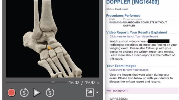
Video radiology reports add value, improve patient care
Radiology reports have traditionally been static text explanations of what is in images, often without links to the images or any layman's explanation of the images for patients. In today's much more visual world of social media, researchers at New York University’s Grossman School of Medicine wanted to see the impact of embedding video into radiology reports to better facilitate patient engagement and understanding of their medical conditions.
The study found it had a big impact and has the potential to improve radiologist communication with patients. Researchers said it highlights the importance of and the ability for imaging to be a part of patient-centered care and that pictures and video are worth a thousand words or more.
The study was published in the American Roentgen Ray Society (ARRS) journal the American Journal of Roentgenology (AJR).[1]
“Patient-centered video radiology reports are a useful tool to help improve patient understanding of imaging results,” explained lead researcher Michael P. Recht, MD, the Louis Marx professor and chair of radiology at New York University’s Grossman School of Medicine, in a statement about the study. “The mechanism of creating the video reports and delivering them to patients can be integrated into existing informatics infrastructure.”
Recht and colleagues collaborated with Visage Imaging GmbH to build an integrated video reporting tool inside the diagnostic viewer, allowing for both image and voice capture. To aid patient understanding of cross-sectional images, cinematic rendered images were automatically created. For their incorporation into video reporting, these images were immediately available at the workstation. Video radiology reports were then uploaded to the institutional health portal, alongside clinical notes and examination images. Finally, a 10-question survey was administered to patients, assessing their perception of the video reports and requesting feedback
From Sept. 20, 2021, to Jan. 22, 2022, 105 out of 227 faculty radiologists created a total of 3,763 video radiology reports. Patients viewed 864 unique videos.
Based on 101 survey respondents, patients rated their overall experience with video radiology reporting a 4.7 out of 5. Specifically, video radiology reports using lay language and annotated images to help improve patients’ understanding of their results. Of the patients who responded to the survey, 91% preferred having both written and video reports together over having written reports alone.
The average time for radiologists to create a video report was between 2.5 and 6.5 minutes.
“Continued development is necessary to further shorten the creation time, so that use of video reports can expand from limited use in selected cases to more widespread use in daily clinical practice,” Recht et al. added in the study.
Find more imaging informatics news
Find more enterprise imaging news
Reference:

