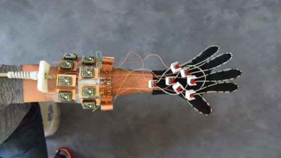
Wearable MRI glove captures clear images of bones, tendons moving together
A novel, flexible MRI component hit the national stage this week in the form of a glove, New York University researchers report. It is delivering science’s first-ever clear images of bones, tendons and ligaments moving together.
A team led by NYU research scientist Bei Zhang, PhD, wrote in Nature Biomedical Engineering that the new MRI element design, which was woven into a cotton glove and tested in a hand playing the piano and picking up objects, has the potential to aid daignosis of repetitive stress injuries, guide surgeries, help build more realistic prosthetics and contribute to scientists’ knowledge of hand anatomy.
“Our results represent the first demonstration of an MRI technology that is both flexible and sensitive enough to capture the complexity of soft-tissue mechanics in the hand,” Zhang said in a release from NYU.
MRI, Zhang and colleagues said, has struggled to overcome limited movement and flexibility. Traditional scanners measure signals that create currents in receiver coils, which are designed to be low-impedance structures and let the current flow freely; the researchers’ new design eliminates electric current altogether, creating a high-impedance structure that blocks current and measures the voltage of magnetic waves as they attempt to establish a current in the coils.
This means the receiver coils can no longer generate magnetic fields that would interfere with neighboring receivers, Zhang et al. wrote, so a rigid structure isn’t necessary any longer. And the results of the first non-rigid prototype, they said, were “exquisite.”
Tendons and ligaments are particularly hard to image since they’re made of such dense proteins—the opposite of what MRI excels at imaging, which includes water-rich soft tissues like muscles, nerves and cartilage. Because of their density, they appear as black bands in scans.
The MRI glove could be an effective method for imaging those dense elements, the researchers said. They were able to observe how the tendons and ligaments, though still appearing as black bands, moved together with bones in the hand.
“We wanted to try our new elements in an application that could never be done with traditional coils, and settled on an attempt to capture images with a glove,” senior author Martijn Cloos, PhD, said in the NYU release. “We hope that this result ushers in a new era of MRI design, perhaps including flexible sleeve arrays around injured knees or comfy beanies to study the developing brains of newborns.”

