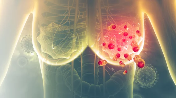
10 clues suggest scope, shape of AI’s future in mammography
Computer-aided detection boosted by AI has often proven superior to traditional CAD over the past decade, yet the “new way” has been slow to win broad adoption.
The lag is largely the result of a high cost of entry combined with knotty ethical and legal concerns. The sooner breast radiology works out the principles and particulars of these issues, the better breast imagers will be at optimizing patient care.
The promise remains worth pursuing, as AI-based CAD tools have been shown to reduce rates of recalls rates and interval cancers while streamlining workflows by automatically de-prioritizing negative mammograms.
So conclude Columbia University researchers who reviewed the literature on AI in mammography and had their report[1] published May 15 in Clinical Imaging.
In summarizing recent developments in AI-aided mammography and considering where momentum points to AI’s future in the field, Richard Ha, MD, and Meghan Jairam, MD, present illustrative findings from more than 30 published studies.
Among the pearls they pull out are 10 key findings in five categories:
Comparison of traditional CAD and CAD based in convolutional neural networks (CNNs)
- When compared to conventional CAD for digital mammography, CNN-assisted CAD has shown a reduction in false positives, improved sensitivity and nearly a 7% increase in area under the curve (AUC).
- Because CNN-CAD systems have lower false positive rates than traditional CAD systems, they may be used to reduce recall rates. In some cases, the recall rate has been reduced by 10 to 20% due to a greater ability to distinguish benign images from malignant ones. This can reduce patient anxiety and distress associated with being recalled.
AI as an assistive tool
- When used as an assistive tool for human readers for digital mammography, the AUC of readers was statistically significantly higher with AI support than without it. With AI support, AUCs ranged from 0.797 to 0.89. Without AI support, these AUCs ranged from 0.769 to 0.87.
- In two of these studies, sensitivity also significantly increased with the use of deep learning. In one study, reading time did not differ when AI support was used. In another, reading time decreased with the use of AI.
Standalone comparison to radiologists
- In some studies, standalone deep learning software statistically significantly outperformed readers. When AUCs for AI programs were obtained in these studies, they ranged from 0.876 to 0.940, when compared with readers’ AUCs, which ranged from 0.778 to 0.81.
- However, in two out of the three studies where AI outperformed readers, the gold standard used was the radiologists' interpretation. In other studies, standalone deep learning was found to have a similar performance when compared to radiologists.
Deep learning for reducing interval cancer rate
- AI programs can be used retrospectively to re-assess … cancers that were correctly marked by CAD as high-risk. In 2019, Hinton et al. showed an increase in AUC for the detection of interval cancer from 0.65 to 0.82 for full-field digital mammography when using AI. In 2021, Lang et al. and Graewingholt et al. showed that deep learning for digital mammography resulted in a 19.3% reduction in interval cancer and had a sensitivity of 48% for identifying interval cancers, respectively.
- [I]n the study conducted by Lang et al., two breast radiologists classified prior mammograms with interval cancers as either true negative, minimal signs or false negative. This type of classification also occurred by radiologists in the study conducted by Graewingholt et al. Hence, one limitation of both studies is that observer bias may have falsely elevated the number of lesions determined by the radiologists to be “false negatives,” which may have inflated the rate of interval cancer detection by the AI software.
Triage screening mammograms
- For digital mammography and DBT, many studies have shown that an AI system can assist with identifying normal mammograms as cancer free, with Yi et al. indicating that deep learning can discard 53% of normal images, which were all found to be cancer free. The deep neural network used by Kyono et al. was able to identify 91% of negative mammograms in a dataset with a cancer prevalence of 1%.
- In addition to reducing radiologist workload and costs related to screening, AI programs can reduce the number of false positives by identifying normal mammograms as negative. This is particularly helpful when the rate of recall is high, such as in the USA, because a reduction in false positives will reduce the number of benign exams that are recalled.
Ha and Jairam also look at research into potential use with AI as a second reader, AI’s technical limitations and potential challenges to wide adoption of mammography AI in the U.S.
Commenting on the latter, they note that, along with costs and ethical and legal hurdles, patient privacy remains a concern.
“Many companies, such as Facebook and Google, that are investing at the intersection of AI and healthcare do not fall under HIPAA, so the health data they collect is not protected under HIPAA,” the authors write. “In the future, these massive amounts of stored health data may be vulnerable to cybersecurity threats as well.”
And yet:
Despite [all] these challenges, AI will likely continue to improve as an assistive tool in breast imaging with a great potential to improve breast cancer screening.”
More Coverage of AI in Breast Imaging:
Malignant architectural distortion ably diagnosed on breast imaging by human-AI combo
AI shows promise as a second reader for breast cancer detection
AI tool increases radiologists’ accuracy at spotting breast cancer on ultrasound scans by 37%
AI-based mammography triage helps providers dramatically improve interpretation process
Pairing ultrasound with artificial intelligence reduces unnecessary breast biopsies
Reference:
Richard Ha, Meghan Jairam, “A review of artificial intelligence in Mammography.” Clinical Imaging, May 15, 2022. DOI: https://doi.org/10.1016/j.clinimag.2022.05.005

