4 ways virtual, augmented reality could change radiology forever
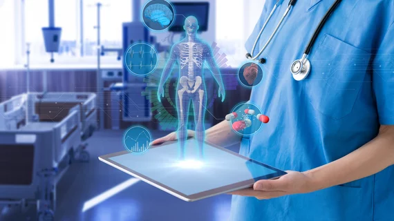
Virtual reality (VR) and augmented reality (AR) technologies have the potential to dramatically change healthcare and could impact the practice of radiology in a number of ways. A team of researchers explored this issue at length, sharing their observations in Radiology.
VR, the authors explained, is when the user is in a completely immersive environment. AR does not involve an immersive environment—instead, the user sees “hologram-like entities” appear in their real environment.
“A number of fields ranging from entertainment and gaming to the military, education, and sports have adopted these technologies for use within their respective fields,” wrote Raul N. Uppot, MD, department of radiology at Massachusetts General Hospital in Boston, and colleagues. “These technologies provide stereoscopic and three-dimensional (3D) immersion within an environment, as in VR, or overlaid onto a real-world background, as in AR. Many medical specialties are now exploring the application of these tools for their respective fields.”
These are four key ways Uppot et al. believe VR and AR can change radiology in the near future:
1. They can improve radiology training
“An important use of VR and AR in medical imaging is to supplement radiology training,” the authors wrote, noting that prior research has found that immersing the learner into a virtual world “is associated with a higher level of active learner participation, owing to increased social, environmental and personal presence within the learning activity.”
AR is already being used to help some trainees “conceptualize complex anatomy,” the authors noted, through the process of converting DICOM images to 3D objects. VR, on the other hand, can help a trainee practice how to react in certain situations such as contrast agent reactions.
Yet another way these technologies can help with training students is by improving how professors give lectures. Uppot and colleagues detailed how their institution has used VR when training interventional radiology students.
“These are interactive lectures that involve students viewing interventional procedure suites using stereoscopic viewers attached to their personal smartphones,” the authors wrote. “These lectures present a case and ask management questions of medical students, including questions about diagnosis, indications, contraindications, type of equipment, and type of sedation. Students then scan a Quick Response (QR) code and are transported to a virtual interventional radiology suite, where they can look around and choose equipment to use for the particular case.”
Applications also exist for training residents, fellows and attending radiologists, the authors added.
2. They can help colleagues communicate with one another
“Planning and executing complex surgical procedures require a comprehensive understanding of anatomic relationships,” the authors wrote. “For the novice proceduralist or surgeon, there are several commercially available simulators available. Three-dimensional learning environments, such as those available using VR technology, offer the potential to increase spatial representation and improve the planning of complex surgical tasks, such as those required of a neurosurgeon.”
Users can also share AR images remotely, according to Uppot and colleagues, allowing them to interact with the same images at the same time from different parts of the world.
3. They can improve communication with patients
VR and AR technologies can help educate patients in a number of ways. For example, at their own institution, the authors have been experimenting with how VR can help patients become comfortable with interventional radiology procedures. They used 360-degree recordings of their interventional radiology suites and then allows patients to explore those areas using VR applications.
“Using stereoscopic viewers attached to smartphones, patients can look around the room and get oriented to the procedure suites and recovery rooms,” they wrote. “Immersive reality can also stimulate a procedure so that a patient can understand the steps of the procedure and what he or she would undergo during the procedure.”
More specific research is needed, the authors wrote, but “radiologists are traditionally early adopters of new technology, so the use of AR and VR technology for patient engagement and education offers interesting potential.”
4. They can provide assistance with interventional radiology procedures
Uppot and colleagues noted that guiding needles, wires and catheters to the targeted area is always a challenge when performing interventional radiology procedures. AR can help with this in a big way.
“An interesting concept that makes use of AR technology is to overlap diagnostic images of a particular lesion directly onto the patient,” the authors wrote. “By overlapping the images on to the patient, the operator can visualize the fused anatomy and proceed with targeting the lesion, while still standing next to the patient. Similarly, AR has been used to provide these types of intraoperative fusion images in open surgery, neurosurgery, and tracheostomy tube placement.”
Of course, there are limitations to these technologies …
As the authors mention near the end of their analysis, VR and AR are still “in their infancy.” In addition, the costs associated with these applications can be quite high and there is still a “limited availability of content.” Still, significant growth is expected in the near future, meaning that some of these limitations could soon be ancient history.
“The clinical utility of both AR and VR in patient education and perioperative planning is promising,” the authors concluded. “Further technical innovations enabling further miniaturization of hardware and greater reliability of registration of images will allow these technologies to redefine procedural planning and patient engagement.”

