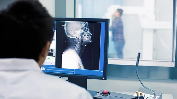
Technology may have been a key contributor to the growing epidemic of burnout among radiologists, but it could also end up being the cure.
Informatics-based changes to imaging tech, when deployed correctly, could end up reducing time spent away from interpreting, while also decreasing interruptions and fueling connections with other clinicians. Experts from Seton Hall University and Penn’s Perelman School of Medicine recently made their pitch for informatics as a burnout-buster in a new analysis, set to be published in February’s Clinical Imaging.
Addressing this surge of workplace fatigue will require organizational changes, they argue, and not simply yoga sessions or reminding employees to take better care of themselves.
“While technological advances have yielded tremendous upsides, they are not always utilized in ways that best serve radiologists,” wrote Andrew Simon, PhD, a psychology professor at Seton Hall, and colleagues. “We suggest that improvements to the workplace can be found through informatics-based strategies.”
Simon and co-authors—who include informatics and radiology experts—spell out seven ways that radiology practices can begin to transform their use of technology:
1) Virtual rounds: Current technology allows radiologists and teams in the intensive care unit to connect using web cameras. Past studies have demonstrated that such interaction boosts ICU providers’ confidence in image interpretations by upward of 90%, while radiologists reported greater understanding of their cases. This could potentially disturb workflow, but authors suggested adopting separate daily rounds dedicated to noninterpretive tasks to help minimize these interruptions.
2) Linking PACS to the EHR: Contacting a referring clinician to convey results can become a big time-waster. The authors suggested linking PACS to the electronic health record to streamline communication and track clinical responsibilities. Providers could adopt a standard template to document important findings, which could be directly imported into the system. Doing so helped decrease turnaround time by more than 4 minutes while also producing “extremely high radiologist satisfaction,” one study found.
3) Deconstructed PACS: Pulling apart your picture archiving and communication system is another option, authors noted. New York University Langone Health Radiology recently tried this method, separating its PACS into six applications, with each component addressing a different duty—interpretation, distribution, storage, etc. NYU has seen progress in improving interoperability and efficiency with this approach. Adopting a PACS-based messaging system to eliminate the need to keep closing out studies is another option, they added.
4) Pop-ups: Informatics can help to improve image interpretation by offering automated EHR searches with relevant clinical or lab information automatically surfacing in a study review window. It could also offer predetermined, relevant info based on the clinical indication or modality, or present a link to a progress note written at the time a referring physician ordered the exam. The latter would make it easier for radiologists to understand the rationale for the study request, Simon noted.
5) Automated dictation process: Further time could be saved by automating parts of the radiologist’s dictation routine, using a program that populates relevant fields in the template, such as contrast agent type and dosing. One study found that such automation saved providers upward of 81 seconds per dictation, or about an hour each work day.
6) Artificial intelligence: There’s no shortage of ways in which AI can help lessen the load on busy radiologists. One example is the use of computer-assisted detection programs to assess lung nodules and pleural effusions. One study found that such programs helped to reduce radiologists’ CT reading times by upward of 44%.
7) Radiomics: This development in nuclear medicine combines technology with informatics advances to allow clinicians to extract quantitative information from anatomical or molecular imaging studies, the authors noted. Technology has now made it possible to correlate radiomic data with biological information and clinical outcomes to help support decision-making. This has allowed radiologists, for instance, to improve tumor control by altering the radiotherapy treatment plan mid-course.
“It is through this type of technological advancement that radiologists have some of their most impactful interactions, guiding the development of precision diagnosis and treatment,” Simon and colleagues wrote. “Such contributions reinforce the value provided by radiologists to those providing clinical care and help reconnect the radiologist with the broader healthcare team.”
Implementing all of these changes will require an investment, and commitment from leadership. But that pales in comparison to the billions in healthcare dollars lost each year to physician turnover and reduced time spent at the bedside, the authors concluded.
“The examples above highlight improvements that do not require a complete overhaul of a system in use. Even small adaptations can be valuable when targeted to the needs of radiologists,” they wrote. “In the end, this will serve not only the radiologist, the healthcare team and the organization, but will produce exponential benefits to patients,” the team added later.

