Ultrasound imaging startup exits ‘stealth’ mode with $4M Department of Defense contract
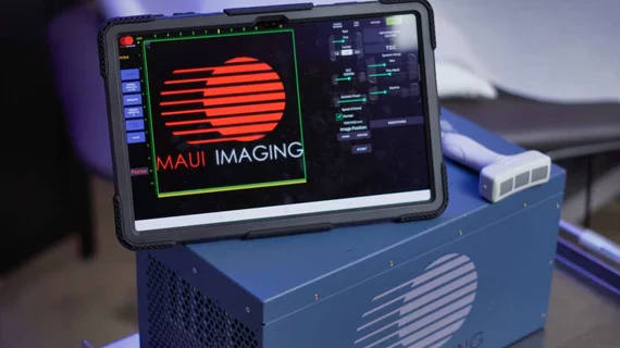
Maui Imaging—a startup offering an ultrasound system that can visualize anatomy others cannot—exited “stealth” mode Thursday, announcing a $4 million contract with the Department of Defense.
Based in Tucson, Arizona, the company’s computed echo tomography product uses sound waves to cross through body barriers for “rapid and effective imaging.” Traditional ultrasound cannot image through the skull without a significant fracture or surgically removing bone. However, Maui Imaging said its product can address this challenge, with impediments such as gas, fat and implants becoming part of the image, rather than obstacles.
The results look like a “cross between ultrasound and CT,” without the need for ionizing radiation. Scans also create a “significantly larger data set,” which can be sliced into additional images and used for artificial intelligence, the company noted.
“The feedback we have received from physicians and technologists highlights the profound need for a new ultrasound-based technology that enables imaging of all types of tissues,” CEO and Co-founder David Specht said in an Aug. 22 announcement. “That need is most pronounced in trauma medicine, which is a major focus of Maui’s collaborative development efforts. Going forward, Maui will be able to supply the volumetric imaging data for AI tools that predominantly come from CT and MRI.”
Specht and colleagues see promise in military medicine, where soldiers often require this level of clinical insight but lack access to advanced imaging. With the seven-figure contract, Maui will support trauma care across the four branches of the military. The company said it hopes to enable faster diagnoses and interventional radiology care in mass-casualty or resource-limited environments.
Maui Imaging’s scientific exploration is supported through the Combat Casualty Care Research Program. Experts will test the technology at the University of Maryland Shock Trauma Center in Baltimore, a major center for training military surgeons. The program hopes to demonstrate that Maui can improve time to care in trauma patients and bolster outcomes. Phase 1 of the project will focus on developing procedures and techniques for using Maui to image 60 different anatomic regions in standardized fashion.
“The ability to rapidly evaluate trauma without the need for immediate access to X-ray, CT and MRI facilities would be of tremendous value to trauma victims worldwide who have sustained significant injury,” Rosemary Kozar, MD, PhD, a University of Maryland professor of surgery, and co-director of the Shock Trauma Anesthesia Research Center, said in the announcement. “Maui could be game changing in a mass casualty setting, underdeveloped countries and on the battlefield.”
The Maui K3900 previously received U.S. Food and Drug Administration clearance in October and is available commercially. Utilizing a concave probe, the device sends and receives energy from multiple angles, allowing it to see “through and around barriers.” Maui also uses proprietary algorithms to accommodate the reflected energy from various flight paths and sums up the data to create a reliable image of structures below the probe. Currently, traditional ultrasound represents less than 10% of AI in medical imaging, but Maui hopes its approach will “dramatically change this.”

