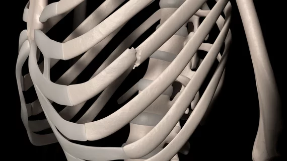
Some patients who suffer blunt-impact injuries of the chest are well served by serial multimodality radiology exams combined with clinical assessment by a trauma surgeon.
The comprehensive approach may be warranted, on a case-by-case basis, for patients whose musculoskeletal injuries include fractures of the costal cartilage. This is the supple connective tissue joining the ribs with the sternum that lets the ribcage expand for breathing.
The conclusion is suggested by findings from a prospective study conducted at Helsinki University Hospital in Finland and published June 4 in Emergency Radiology [1].
Noting that the most common cause of costal cartilage fracture (CCFX) is vehicular collision, lead author Mari Nummela, MD, and colleagues point out that these fractures are not visible on plain X-rays and can be difficult to discern alongside fractured ribs.
In such cases, ultrasound alone may reveal a costal cartilage fracture, but cross-sectional imaging with CT and MRI “is essential to verify the clinical suspicion of CCFX,” the authors write.
It’s important to pin down the CCFX diagnosis, they add, because fractured cartilage “contributes to ribcage instability and may present as prolonged post-traumatic pain and discomfort even months after the initial trauma.”
For their study, Nummela and colleagues, including senior author Seppo Koskinen, MD, PhD, of the Karolinska Institute in Sweden, examined 21 patients diagnosed with CCFX in the ED.
The team sent all 21 for serial MRI, ultrasound and ultra-low-dose CT at initial presentation and in follow-up exams at an average of three years after the trauma.
The patients also were seen over time by an orthopedic specialist, who largely focused on pain and mobility levels.
On review, the costal cartilage fractures were deemed stable by radiologists in 71.4% of the patients (n = 15) at follow-up.
In addition:
- CT showed all patients had calcifications of healed costal cartilage.
- Dynamic ultrasound revealed increased fracture dislocation and persistent movement in five patients (23.8%), while one patient (4.8%) showed signs of a non-stable union.
- MRI was best suited for visualizing local persistent edema.
In clinical follow-ups, four patients (19.0%) reported experiencing persistent CCFX symptoms.
In their discussion, Nummela et al. comment that, in general, most costal cartilage fractures will heal over time and become asymptomatic.
“However, some patients report pain, discomfort and a snapping sensation around the CCFX even years after the initial trauma,” they write. “A diagnostic work-up that consists of both clinical examination and imaging with CT or MRI ensures a comprehensive evaluation.”
More:
Most, but not all, fractures that were radiologically evaluated as non-healed were symptomatic. Conversely, some symptomatic fractures were evaluated as healed on imaging. Thus, the combination of clinical examination and imaging will help assess the overall situation.”
As costal cartilage injuries are commonly seen in high-energy trauma, “other concomitant injuries add to the pain and disability load,” the authors warn, “and cartilage injuries may remain overlooked as the source of pain and discomfort.”
The study is available in full for free.
More Coverage of Musculoskeletal (MSK) Imaging:
Wide variation in musculoskeletal imaging charges, including 74-fold difference for one CT exam
Radiologists able to flag domestic violence before victims self-report
Orthopedic surgeons share what the specialty seeks in a quality radiology report
Orthopedic surgeons’ most common reasons for opting not to read radiology reports
Reference:
- Mari Nummela, Seppo Koskinen, et al., “Costal cartilage fractures in blunt polytrauma patients—a prospective clinical and radiological follow-up study.” Emergency Radiology, June 4, 2022. DOI: https://doi.org/10.1007/s10140-022-02066-w

