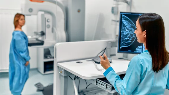
How 1 radiology department cut its mammography turnaround times by 39%
An academic imaging department has cut its mammography turnaround times by nearly 39% while also significantly reducing radiologist fatigue, according to new research published Thursday.
Amid the COVID-19 pandemic, Michigan Medicine saw its peak processing of breast images climb to 198 hours or about 8.25 days. Leaders identified three major drivers of this figure—radiologist interruptions, paper-based workflows and “cumbersome” report dictation processes.
To begin addressing this lag, Michigan Medicine developed a new “batched, digitized” workflow using a reporting aid they call “uninterrupted with assistant,” experts detailed in Current Problems in Diagnostic Radiology. The practice change has produced dramatic gains, with average turnaround times falling from over 83 hours down to 51 after the project.
“These interventions offer strategies to improve productivity and address practical issues of burnout and workforce retention,” lead author W. Tania Rahman, MD, and colleagues wrote Jan. 23.
At the height of Michigan Medicine’s imaging backlog, radiologists were reading images in an “interrupted fashion,” often between procedures and diagnostic cases. Work was handled via an “unintegrated,” vendor-specific PACS utilizing nonstandardized report templates. Physicians were required to navigate through a series of mouse clicks and drop-down menus to transfer the BI-RADS (Breast Imaging Reporting and Data System) code and management recommendations into the electronic medical record.
To address this problem, Michigan Medicine first shifted to a new batch-reading approach where radiologists read groups of mammograms uninterrupted and free from competing tasks. The organization deployed one clerical staff member as a “live transcriptionist,” sitting alongside physicians to review charts and draft pre-dictated reports in “real time.” Under this new “uninterrupted with assistant” arrangement, radiologists typically took on four-hour reading shifts, tackling an “equitably assigned” work list of screening mammograms.
Next, Michigan Medicine shifted toward digitizing workflows and eliminating the use of paper in its processes. Patients were asked to complete intake forms on a tablet, and technologists were trained to enter data on a computer. Paper documents were scanned and uploaded to the EMR, while paperwork to communicate diagnostic recall views were replaced with a standard electronic protocol. Finally, the institution moved toward a standard mammography template with pre-populated, modifiable fields. These utilized U.S. Food and Drug Administration-approved verbiage to comply with audit measures. Drop-down menus were replaced with pre-filled fields for a “more effortless experience,” the authors noted. However, image viewing remained unintegrated with the EMR and dictation software.
Rahman and colleagues tracked a 17-week preintervention period spanning until Jan. 31, 2020, when radiologists worked in their interrupted eight-hour reading shifts. They compared this up against a 15-week post-intervention period spanning until July 3, 2021. A total of 14 fellowship-trained breast imaging radiologists with varying levels of clinical effort took part in the study. Michigan Medicine aimed for an institutional turnaround time of less than 72 hours after the practice changes. Researchers also administered radiologist surveys to help gauge fatigue and distraction levels on a 10-point scale.
Prior to the changes, only about 35% of the rads met their benchmark of report turnaround times shorter than three days. Afterward, this number leapt to 93%. Average weekly volume of screening mammograms was “significantly” lower after the intervention, potentially impacting the findings. But when controlling for various factors, average weekly reported turnaround times remained much lower following the changes. Radiologist interruptions and fatigue also dropped markedly using the new approach, the authors added.
“Future improvements include transitioning from a digital workflow to an integrated system with communication between the image viewing system, radiology information system and reporting software, which can further improve efficiency and accuracy,” the authors noted. “Such a system can be compatible with remote workstations and predrafted reports that the transcriptionist can generate independently. This may create greater workplace flexibility and job satisfaction.”

