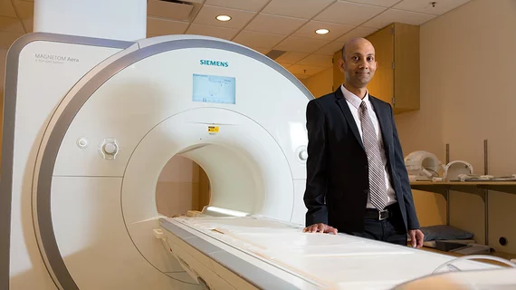
First-of-its-kind study proves cardiac MRI ‘powerful’ for assessing suspected heart tumors
A first-of-its kind study is proving that cardiac MRI is a powerful predictor when assessing heart tumors and should serve as the “technique of choice” for physicians.
In an analysis spanning four centers and more than 900 patients, scientists discovered that cardiovascular magnetic resonance (CMR) diagnoses were 98.4% accurate. Lead author Chetan Shenoy, MBBS, labeled this an important study because there is little written on the use of MRI to assess such patients.
“These data provide the first large-scale validation of the clinical practice of using CMR to exclude a cardiac tumor and to diagnose the type of tumor,” Shenoy, an associate professor and cardiologist with the University of Minnesota Medical Center, said Oct. 7. “By demonstrating high accuracy and long-term prognostic value, our results confirm that CMR can be used as a ‘one-stop-shop’ for this clinical indication. We anticipate our findings will shape future guidelines, appropriateness documents and health policies on this topic.”
The analysis included patients undergoing an MRI for suspected cardiac tumors. Among a total of 903 patients, 25% had no mass, 16% a pseudomass, 16% a blood clot, 17% a benign tumor and 23% were of the malignant variety. After a median span of nearly five years, 376 patients died (the study’s primary end point was all-cause mortality). When comparing up against the final diagnosis, CMR was accurate for 98.4% of patients. Those with no mass, a pseudomass or benign tumor all had similar long-term death rates. The technique additionally provided “incremental prognostic value” over other clinical factors such as left ventricular ejection fraction, coronary artery disease, and a history of extra-cardiac malignancy
“The CMR diagnosis is a powerful independent predictor of mortality incremental to clinical risk factors,” Shenoy and colleagues concluded Sept. 21 in the European Heart Journal.

