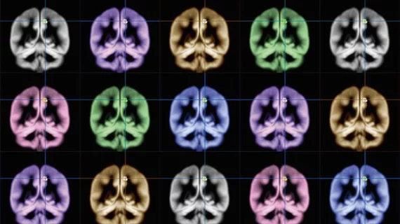
MRI-compatible tech allows for imaging without contrast agents
MRI-compatible technology out of Purdue University has the ability to detect and monitor cerebral vascular disorders and injuries without the use of potentially harmful contrast agents, the university announced this week.
Rather than focusing on traditional imaging techniques, which involve injecting patients with agents like gadolinium to calculate cerebral circulation, a team led by Yunji Tong, PhD, opted for a method that tracked intrinsic blood-related MRI signals that travel with a patient’s blood flow.
“We can compare the signal from symmetric arteries and veins in both hemispheres or neck to assess the cerebrovascular integrity, or the balance of blood flow,” Tong, an assistant professor of biomedical engineering at Purdue, said in a release. “The blood flow should be symmetric between the two sides in a healthy subject.”
Tong et al. calculated cerebral circulation time—the delay between intrinsic signals from the internal carotid artery and the internal jugular vein—using this alternate method, which can indicate blood flow disturbances, tumors, traumatic brain injuries or other brain diseases by measuring time intervals. That calculation is possible with conventional imaging, the researchers said, but only using contrast agents, and only for a few seconds at a time after the patient has been injected.
The Purdue team’s method, on the other hand, allowed for continuous monitoring of the circulation time.
“The new method can even be applied on some existing MRI data to calculate the cerebral circulation time,” Tong said. “This method is safer and non-invasive since we don’t inject contrast agents, which can stick to vessels or cause other health problems.”
Tong’s work was funded in part by the National Institutes of Health and has been published in the Journal of Cerebral Blood Flow and Metabolism.

