Could this research help prove cutting-edge MRI techniques are safe for patients?
Researchers have simulated how more than 20 different breast tissue ratios respond to heat emitted from MRIs at higher field strengths than those currently in hospitals, according to findings in Magnetic Resonance in Medicine.
The researchers, led by Joseph V. Rispoli, PhD, of the Weldon School of Biomedical Engineering at Purdue University in West Lafayette, Indiana, sought to determine how much radiofrequency energy each breast tissue ratio could handle, while remaining within FDA safety limits.
"We're starting to develop techniques at high field strengths that could immediately monitor how tumors respond to treatment,” Rispoli said in a prepared statement issued by the university. “So we don't want tissue heating concerns to stand in the way of improving such a powerful tool."
The density of a woman’s breast determines the amount of radiofrequency energy from an MRI it will absorb in the form of heat, which is defined as the specific absorption rate (SAR). Denser breasts have higher SARs, and it is more difficult to determine the amount of heat that is safe for women.
At present, MRI field strengths are currently up to 3 tesla. The researchers noted, however, many techniques would be “far superior” at 7 tesla. While 7 tesla would come at a five-fold increase in SAR, it would also double the MRI sensitivity, allowing for a better reading.
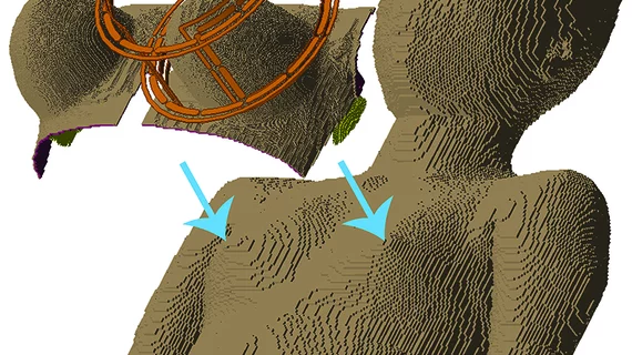
The researchers used phantoms of the female body for their study. Specifically, they fused 38 breast phantoms at various densities as classified by the American College of Radiology Breast Imaging Reporting and Data System Atlas, along with Swiss- and Japanese-created models.
They simulated each fused phantom in response to MRI coils at 7 tesla. Even at 7 tesla, dense breast tissue did not overheat. The simulations, the researchers noted, will help tailor the amount of radiofrequency used for various breast densities.
"We want to facilitate the most cutting-edge of breast MRI techniques at any site in the world," Rispoli concluded. "Ultimately, a woman will be able to go in, have a fast low-power anatomical MRI scan, and then the computer could quickly simulate on-the-fly what the SAR would be in that patient."

