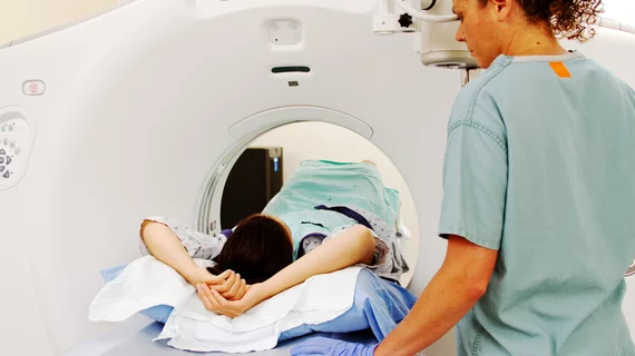
Abdominal MRI intervention helps drop respiratory motion and image quality degradation
An intervention prior to abdominal MRI has helped NYU Langone Health drop respiratory motion and image quality degradation during exams, according to new research.
Technologists can face challenges getting patients to comply during their scans, which is only exacerbated if the subject speaks a different language and cannot comprehend instructions. However, the New York City institution has developed instructional videos in the patient’s primary language to improve adherence during magnetic resonance imaging.
Testing the videos on 29 patients who speak Spanish or Mandarin-Chinese as their primary language, NYU produced promising results. When patients requiring translators viewed the video, T2-weighted image quality improved significantly compared to those who didn’t, experts detailed Thursday in the Journal of the American College of Radiology.
“Radiology departments should strongly consider creating patient-centric abdominal MRI instructional videos in the languages of their most commonly encountered non-English-speaking patients,” Myles Taffel, MD, an associate professor at the Grossman School of Medicine and associate section chief in abdominal imaging, and co-authors concluded.
Currently, more than 60 million individuals in the U.S. speak a foreign language at home, according to census estimates, with 25 million indicating struggles interacting in English. Patients with limited proficiency are at increased risk for poor health outcomes, and language barriers can pose problems in the MRI suite, where abdominal exams come with carefully choreographed breathing instructions.
NYU first created the 2.5-minute videos in October 2019 as part of a quality improvement project at three urban outpatient imaging facilities. Patients who required a translator watched the clip in the prep room prior to undergoing an upper abdominal MRI. Taffel et al. retrospectively analyzed data from 29 patients who did and 50 who did not view the intervention between 2019-2020, also comparing them against 81 English-speaking patients’ results.
Three radiologists independently assessed respiratory motion and image quality on both turbo spin-echo T2-weighted images and post-contrast T1-weighted images using a 1-5 scale. Physicians observed marked motion and image quality degradation on T2WI for those who required a translator, but significant improvement for those who took part in the intervention. Meanwhile there was no notable difference in respiratory motion or image quality for T1WI, with rapid acquisition techniques achieving robust image quality across all patient groups.
The study was limited by its small sample size and other factors. But researchers urged others to test this intervention, underlining the ease of creating such content on smartphones.
“The U.S. healthcare system continues to strive toward the delivery of high-value, high-quality care to all patients equitably, and this mission extends to radiology,” Taffel et al. wrote. “In the setting of a growing population with limited English proficiency, radiology must adapt to address healthcare disparities.”

