AI helps radiologists, ED providers quickly locate coronavirus pneumonia from chest x-rays
When COVID-19 patients develop severe pneumonia, they typically require intensive care, breathing assistance from ventilators, and other scarce resources. Reaching that pneumonia diagnosis early is crucial, but doing so is often a subtle art. One California provider, however, has started deploying artificial intelligence to simplify and speed the process up.
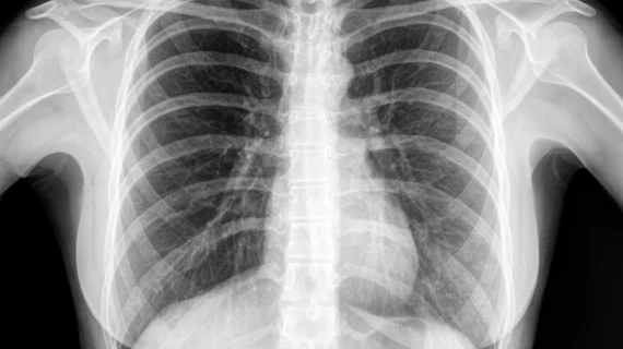
UC San Diego Health radiologists helped develop the AI algorithm that can quickly locate pneumonia from coronavirus patients’ chest x-rays. In one investigation highlighted recently in the Journal of Thoracic Imaging, the algorithm was able to consistently find areas of inflammation and fluid buildup in 10 chest images, gathered from patients treated at different healthcare institutions.
“As we prepare for a potential surge in patients with COVID-19, it’s not just patient rooms and supplies that may become limited, but also physician and staff capacity. So it’s tremendously helpful to have tools that allow physicians who are not as experienced as radiologists in reading x-rays to get a quick idea of what they’re looking at, especially frontline emergency and hospital-based physicians,” Christopher Longhurst, MD, chief information officer and associate chief medical officer, said in a statement.
Radiology professor Albert Hsiao, MD, PhD, and colleagues first devised the algorithm months before the pandemic as a way to detect regular pneumonia from chest x-rays. The team trained the system using 22,000 notations by human radiologists, overlaying images with color-coded maps to show areas of possible pneumonia.
Now, Hsiao and his team are applying the approach to coronavirus cases and finding early success. In one instance, a patient presented at the ED with no COVID-19 symptoms and underwent chest x-rays for other reasons. The AI tool indicated early signs of pneumonia, however, which a radiologist confirmed, and testing eventually led to the coronavirus diagnosis.
“We would not have had reason to treat that patient as a suspected COVID-19 case or test for it, if it weren’t for the AI,” Longhurst said. “While still investigational, the system is already affecting clinical management of patients.”
Hsiao said they’ve focused on chest x-rays rather than CT or other tools, as they’re cheaper, equipment is portable, and results arrive quickly. He emphasized that they’re not actually diagnosing coronavirus itself with the AI system, and results are preliminary. They next plan to expand the investigation to UC San Diego’s four other academic medical centers.

