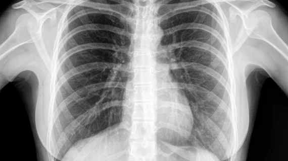
Common features of missed lung cancer on chest X-ray
There are a few common features of missed lung cancer cases on chest X-ray images, according to a new analysis published Tuesday.
Those can include some radiographic features pertaining to the tumor’s location, along with density and any superimposed anatomical structures, experts wrote in Clinical Imaging [1]. The findings are based on an analysis of 75 patients imaged at a single institution, with providers overlooking the initial signs of lung cancer.
Researchers hope the findings will help radiologists to avoid such errors, which can lead to worse patient outcomes and death.
“It is, therefore, crucial for radiologists and clinicians to beware of [missed lung cancer] in routine clinical practice,” corresponding author Thitiporn Suwatanapongched, with the faculty of medicine at Ramathibodi Hospital in Bangkok, Thailand, and colleagues concluded. “Although radiologist performance cannot be perfect and some errors are inevitable, approaches exist to minimize error causes.”
Those can include comparing the chest radiograph being read with several previous examples, focusing on common blind spots, and avoiding mistaking lesions for normal structures. Computer-aided detection systems or artificial intelligence also can enhance the radiologist’s performance and help detect smaller-sized tumors, the authors advised.
For the study, researchers retrospectively reviewed chest X-rays from 95 patients, obtained six months or more prior to a lung cancer diagnosis at their hospital. The final sample included 75 patients (or 79% of the total), evenly split between men and women at an average age of nearly 65 years old.
The median missed lung cancer size was about 16 mm, Suwatanapongched et al. reported. Nearly 75% occurred in the outer two-thirds of the lung, followed by 68% in the middle/lower zones and 55% in the left lung. Common features included anatomical superimposition (in 88% of cases), a density less than the aortic knob (85%), partly/poorly defined margin (77%), an irregular/spiculated border (61%), and a rounded/oval shape (57%). Of the missed cases, almost 47% had stage 3 or 4 lung cancer and 41% of patients died.
“In our study, [missed lung cancer] in the inner one-third of the lung, exhibiting a density equal to/greater than the aortic knob, or superimposed by the midline structures (the hilum, heart and thoracic aorta) was significantly associated with stage 3 and 4 LC at diagnosis,” the authors advised. “The three-year all-cause mortality significantly increased when [missed lung cancer] was in the upper zone, superimposed by pulmonary vessels, superimposed by pulmonary vessels plus ribs, or superimposed pulmonary vessels plus in the inner one-third of the lung. To our knowledge, such associations have never been addressed.”
The authors speculated that density greater than or equal to the aortic knob could signal a more solid or invasive case of lung cancer. More complex anatomy in the inner one-third of the lung and superimposed anatomical structures, meanwhile, might hinder detection, according to the study.
Read more about their findings in the official journal of the New York Roentgen Society below.

