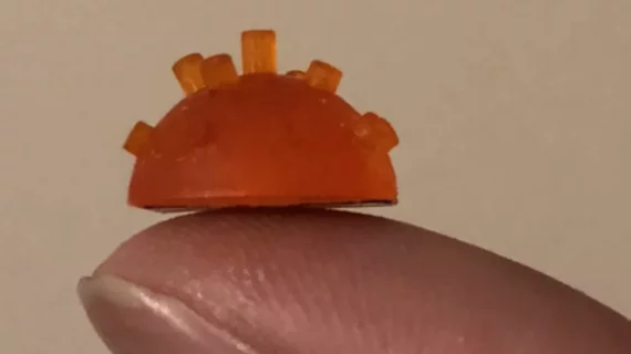
Pipe organ-like ultrasound transducer offers improved quality for medical images
Researchers from the University of Strathclyde in Glasgow, Scotland, have developed a miniaturized version of a musical pipe organ, or a piezoelectric micromachined ultrasonic transducer (PMUT), that could potentially improve the quality of medical images.
Led by Tony Mulholland, PhD, the team studied the efficacy of the PMUT and published their results in the journal IEEE Transactions on Ultrasonics, Ferroelectrics and Frequency Control.
They found the bandwidth of the PMUT is 56 percent and 59 percent in transmitting and receiving ultrasound signals—five times greater than the standard device—which will lead to higher quality medical images.
“Around 20 percent of medical scans are performed using ultrasound,” Mulholland said in a prepared statement. “The scanner creates images by emitting sound waves with a frequency that lies above human hearing. The scanner operates at a single frequency—similar to a piano that can play just one note—and this accounts in part for the relatively poor resolution that one sees in ultrasound images.”
Mulholland and colleagues developed the PMUT using a 3D printer. The idea behind the PMUT being fashioned as a miniature pipe organ is that it can emit sound waves in various frequencies, which Mulholland noted “would provide a marked improvement in the imaging capability.”
“Musical instruments create sounds over a broad range of frequencies and have been carefully designed over the centuries to be very efficient at doing so,” added James Windmill, PhD, professor at the University of Strathclyde, in the same statement. “It is well known that the highest frequency pipes are the smallest in length, as in, for example, a piccolo, so to realize frequencies that are beyond human hearing—ultrasound waves—the length has to be very small indeed, of millimeters in length.”
The researchers tested their designs and found the bandwidth of the PMUT was able to transmit and receive ultrasound signals, five times more than the standard device.
“Developing wide bandwidth ultrasound systems could give significant improvements in imaging capability,” Windmill said. “Using high resolution 3D printers allows us to try new, three-dimensional device designs with much faster development cycles.”
The PMUT is still in the earliest stages of development, though it may impact the design of hearing aids and also “in underwater sonar and the non-destructive testing of safety critical structures such as nuclear plants.”

