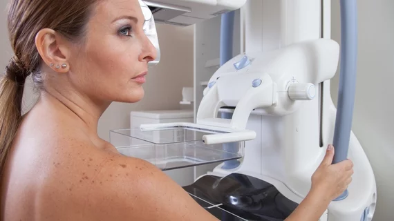
Automated measurements confirm women with dense breasts see higher cancer rates
Women with dense breasts see higher cancer rates than their non-dense breast counterparts, suggesting additional screening might be beneficial, a study from Norway has found.
The research, led by Solveig Hofvind, PhD, of the Cancer Registry of Norway, backs up previous work that’s found higher breast cancer rates in the dense breast population. But while past studies have based results on subjective density assessments, likely produced by radiologists using the American College of Radiology’s Breast Imaging Reporting and Data System, Hofvind’s research uses more streamlined, automated volumetric measurements.
Hofvind et al. used automated software to classify breast density in 107,949 middle-aged women, collecting 307,015 digital screening exams from the patients between 2007 and 2015. The software classified 28 percent of all cases as dense.
“The odds of screen-detected and interval breast cancer were substantially higher for women with mammographically dense versus fatty breasts in BreastScreen Norway,” Hofvind said in a release from the Radiological Society of North America. “We also found substantially higher rates of recalls and biopsies among women with mammographically dense breast tissue.”
The rate of screen-detected cancer in women with dense breast tissue was 6.7 per 1,000 examinations, compared to 5.5 per 1,000 in women with fattier tissue. The former group also saw a 3.6 percent recall rate, while the latter’s was 2.7 percent. Biopsy rates were similar—1.4 percent in the dense breast group and 1.1 percent in the non-dense cohort.
Hovfind said mammographic sensitivity was also significantly reduced women with dense breasts. Whereas sensitivity in patients with non-dense breasts reached 82 percent, it topped out at 71 percent in dense breast patients. Cancers detected in the dense breast group were more advanced and typically larger.
To offset those differences, Hovfind said, it might be helpful for women to be informed about their individual breast densities, hiked risk and any additional imaging options. The superimposition of dense breast tissue on a mammogram can mask areas of the scan, making it harder to detect some cancers.
“We need well-planned and high-quality studies that can give evidence about the cost-effectiveness of more frequent screening, other screening tools such as tomosynthesis and/or the use of additional screening tools like MRI and ultrasound for women with dense breasts,” she said. “Further, we need studies on automated measurement tools for mammographic density to ensure their validity.”
Hovfind and her colleagues’ work has been published in Radiology.

