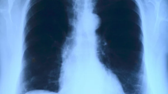
Chest tomosynthesis can accurately ID pulmonary nodule growth, replace CT
Chest tomosynthesis can accurately detect pulmonary nodule growth, according to a new study in Academic Radiology, but clinicians must be aware of certain factors that may influence its accuracy.
“The present study indicates that chest tomosynthesis can be used to detect pulmonary nodule growth,” wrote lead author Magnus Båth, PhD, with the University of Gothenburg in Sweden, and colleagues. “However, nodule size, dose level, and mismatch in position relative to the image reconstruction plane in the baseline and follow-up examination may affect the precision.”
Researchers created a total of 40 simulated baseline nodules with volumes that ranged from 100 mm3 to 300 mm3—the sizes recommended for surveillance in a current low dose CT lung cancer screening trial. Additional versions of nodules were produced for the study. Those with growth volumes ranging from 11 percent to 252 percent were included.
Nodules were inserted into 20 chest tomosynthesis images to simulate a situation where the growth remained stable in size or increased between the two imaging studies. They found tomosynthesis on par with the traditional CT method.
“In general, the possibility of adding a tomosynthesis examination to a chest radiograph in order to rule out or confirm suspicious lesions, and thereby possibly avoiding the need for CT, has been identified as one of the major areas of clinical use for the technique,” Båth and colleagues wrote.
The team also set out to determine how nodule size and dose level affected the accuracy of chest tomosynthesis through an observer study which involved four thoracic radiologists using an area under the curve scoring system.
While the observers could pick out which nodules were stable over time versus those that showed “clinically relevant” growth, authors found the lowest values were present in cases with a mismatch in nodule position relative to the reconstructed image plane between two exams. Overall, they noted nodule size and dose level affected the precision of tomosynthesis.
“These results indicate that, for a given relative nodule volume increase, the detection rate of nodule growth will decrease with the nodule size but that the effect would decrease with increasing nodule growth,” Båth et al wrote.

