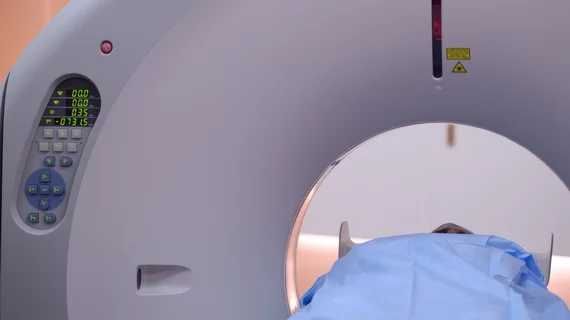
3D camera guidance meaningfully aids patient positioning, dose reduction
Regardless of a patient’s body mass index, using a scanner-integrated 3D camera for situating CT patients on the table can optimize not only patient positioning but also radiation dose, according to researchers in Germany.
Panagiota Manava, MD, of Paracelsus Medical University in Nuremberg and colleagues report their findings in the February edition of Investigative Radiology [1].
The team worked with experienced CT technologists as they imaged around 3,100 patients for chest and/or abdomen studies on a 128-slice scanner outfitted with a 3D positioning camera (Siemens Healthineers’ Somatom X.cite).
They split the patients about evenly into two groups. One group was positioned with conventional laser guidance and the camera switched off. The other group was positioned with guidance from the 3D camera.
The researchers compared positioning accuracy relative to the scanner’s isocenter—the spatial sweet spot at the center of the gantry bore—and analyzed radiation parameters.
Manava and co-authors report isocenter positioning improved significantly with guidance from the 3D camera as compared with visual laser guidance.
They also found absolute table height differed significantly between the two guidance options: The bed was higher with 3D camera positioning (165.6 ± 16.2 mm) as compared with laser-guided positioning (170.0 ± 20.4 mm).
On this aspect the authors point out that laser lines projected on patients help adjust table height in all clinical CT scanners. However, “visual positioning still is user dependent, and even for experienced [technologists], it is not always trivial,” they write.
Closer to the objective of the project, the team found radiation exposure decreased using the 3D camera as indicated by dose length product, effective dose and CT dose index.
Further, exposure to radiation-sensitive organs such as the colon and red bone marrow were lower using the camera.
Commenting on their selection of only experienced and highly trained technologists for the study, the team acknowledges this may represent a limitation in study design.
However, they remark, “the benefit of a camera system may be even greater when less experienced staff is involved.”
Implementation of a 3D camera “improves and homogenizes patient positioning for common CT examinations (chest, abdomen and pelvis),” Manava et al. conclude. “Although most of our patients were positioned within a range of ±4 cm by experienced [technologists], the camera-based algorithm positioned all patients within that range with a much lower standard deviation.”
More:
Correct positioning in the isocenter improves the accuracy of automatic exposure control and automated tube voltage (kVp) selection and also results in lower radiation exposure, independently from the BMI.”
The study is available in full for free.

