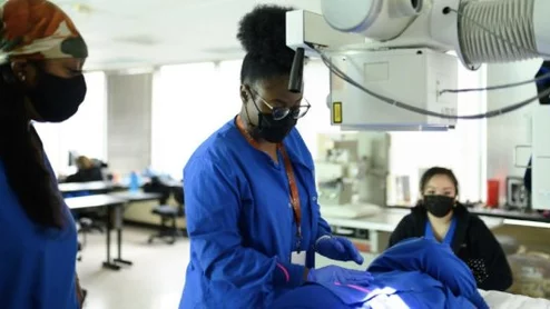
Understanding imaging is beneficial to all medical residents, not just those pursuing a career in the specialty, but many have reported feeling uninformed about the field as a whole.
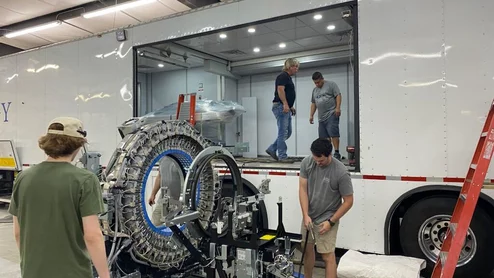
The systems' deaeration hoses in the X-ray tube cooling units may be vulnerable to degrading over time, preventing them from properly cooling and leading to oil leaks.
Based in Mountain View, California, and founded in 2007, Heartflow had originally hoped to raise about $100 million from the IPO, but the tally ballooned amid sizable interest.
The American Society of Nuclear Cardiology visited Capitol Hill to advocate for a variety of issues. Cardiovascular Business spoke to the group's president to learn more.