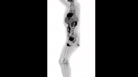
Images from world’s first total-body medical scanner headed to RSNA 2018
EXPLORER, a new total-body PET/CT scanner, is the world’s first medical imaging equipment to produce 3D pictures of the entire human body—and its initial images are headed to RSNA 2018 in Chicago.
The scanner is the work of researchers Simon Cherry, PhD, and Ramsey Badawi, PhD, of the University of California, Davis, and Shanghai, China-based United Imaging Healthcare. Cherry and Badawi first had the idea for EXPLORER 13 years ago, receiving a $1.5 million grant from the National Cancer Institute to get started back in 2011. In 2015, the team received $15.5 million from the National Institutes of Health.
EXPLORER can capture scans in just one second and produce movies that track how certain drugs move through a patient’s body. It also exposes patients to up to 40-times less radiation than modern PET scans.
“While I had imagined what the images would look like for years, nothing prepared me for the incredible detail we could see on that first scan,” Cherry, a distinguished professor in the UC Davis department of biomedical engineering, said in a prepared statement from the university. “While there is still a lot of careful analysis to do, I think we already know that EXPLORER is delivering roughly what we had promised.
“The level of detail was astonishing, especially once we got the reconstruction method a bit more optimized,” Badawi, chief of nuclear medicine at UC Davis Health, said in the same statement. “We could see features that you just don’t see on regular PET scans.

