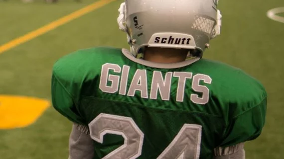
RSNA 2018: MRI scans reveal what football does to young athletes’ brains
Repeated blows to the head can cause changes to the brains of young football players, according to a new study presented at RSNA 2018 in Chicago.
“The years from age 9 to 12 are very important when it comes to brain development,” study lead author Jeongchul Kim, PhD, from Wake Forest School of Medicine in Winston-Salem, North Carolina, said in a prepared statement. “The functional regions of the brain are starting to integrate with one another, and players exposed to repetitive brain injuries, even if the amount of impact is small, could be at risk.”
Kim’s team examined MRI scans from 26 male football players before and after playing three months of football. The players had an average age of 12. Twenty-two boys with similar ages who did not play contact sports also underwent MRI scans at the same time so the authors could make comparisons.
Overall, the scans revealed the football players had changes in their corpus callosum, “a critically important band of nerve fibers that connects the two halves of the brain,” according to the study. Signs of axial strain and radial strain were observed by Kim and colleagues.
“The body of the corpus callosum is a unique structure that's somewhat like a bridge connecting the left and right hemispheres of brain,” Kim said. “When it's subjected to external forces, some areas will contract and others will expand, just like when a bridge is twisting in the wind.”
The team hopes its research can help with the development of future guidelines that ensure young athletes are as safe as possible when playing football.
The authors of a recent study published in Neurobiology of Disease also reported observing changes in the brains of athletes due to playing football. Additional Radiology Business coverage of this topic can be read here and here. Similar findings to this study were reported in another recent study

