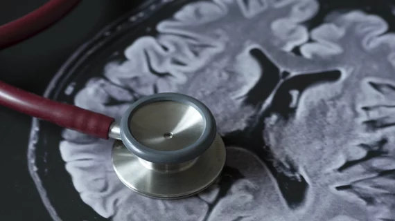
AI helps bolster radiologists’ ability to detect ADHD using MRI
Radiologists have discovered a way to boost their ability to detect attention deficit hyperactivity disorder (ADHD) using deep learning-aided MRI, a development that could aid in pinpointing other neurological conditions.
Cincinnati researchers recently made this breakthrough using functional magnetic resonance imaging (fMRI) to create comprehensive maps of the connections between brain networks. Such “connectomes,” as they’re called, could be key to understanding difficult-to-diagnose brain disorders such as ADHD, they noted in their analysis, published Wednesday, Dec. 11, in Radiology: Artificial Intelligence.
The team was able to build a deep learning model using multi-scale brain connectome data from nearly 1,000 participants, coupled with personal characteristics like gender and IQ. The model was able to “significantly” increase ADHD detection when compared to the single-scale method, constructed using only one parcellation of the mind.
“Our results emphasize the predictive power of the brain connectome," senior author Lili He, PhD, with Cincinnati Children's Hospital Medical Center, said in a statement.
This work is crucial, He and colleagues noted, with about 6.1 million kids (or 9.4%) diagnosed with ADHD each year. Doing so has proven difficult in the past, with clinicians relying on several symptoms and behavioral tests, rather than a single imaging exam.
Scientists believe the disorder stems from a breakdown or disruption in the connectome, and He and colleagues hope to eventually explore the root causes behind these brain hiccups. They’re hoping that spotting these markers using MRI might aid in diagnosis of ADHD earlier in childhood, before kids begin falling behind their peers.
“This model can be generalized to other neurological deficiencies," He added, noting that it’s already used to predict cognitive deficiency in pre-term infants.

