Letting Deep Learning Light the Way to Translational Research and Precision Medicine
Quantitative traits obtained from CT scans performed in routine clinical practice have the potential to enhance translational research and genomic discovery when linked to electronic health record (EHR) and genomic data. For example, both liver fat and abdominal adipose mass are highly relevant to human disease: Non-alcoholic fatty liver disease (NAFLD) is present in 30% of the U.S. adult population, is strongly associated with obesity, and can progress to hepatic inflammation, cirrhosis and hepatocellular carcinoma. These appear as incidental findings on CT, but the clinical significance is unclear and needs referral to a hepatologist. The automated extraction of liver fat and adipose mass from clinical imaging studies at scale would be of great interest for integration with other aspects of the phenome via the EHR, as well as potentially with genomic and biomarker data, to advance translational science and precision medicine and become a value-added feature of a radiology report.
Aims and objectives. Our primary pursuit was developing a fast, fully-automated, cloud-based AI medical image processing pipeline for near real-time segmentation, feature extraction and reporting on clinical liver CT images from PACS. At the same time, we wanted to quantify hepatic fat from abdominal CT studies and associate it with genetic and phenotypic markers in the Penn Medicine Biobank (PMBB).
Leadership and project management. Walter Witschey, PhD, assistant professor of radiology at Penn Medicine, led the project. His team included Mathew MacLean, MD, who led the software development and implementation while as a final year med-student at Penn. They worked closely with Rotonya Carr, MD, a hepatologist, and Dan Rader, MD, a professor of genetics and medicine who heads the Penn Biobank.

Key steps. Using deep learning, we automatically extracted 12 quantitative imaging traits from more than 150,000 chest and abdominal imaging scans from 19,624 adults. In a phenome-wide association study, we compared these traits with diagnostic codes and found 2,495 significant associations with the disease phenome. This work leverages the Penn Medicine BioBank (PMBB), which is a unique and powerful resource for this investigation and has obtained genetic and blood biomarker data for these patients, representing the largest and most diverse cross-section of Americans to date.
Positive outcomes. 1.) Going by total number of imaging scans analyzed, quantitative traits extracted, and association with genetic and other phenotyping data, our study is the largest of its kind. By comparison, a study published last year analyzed one trait (liver fat) in less than half the number of patients and did not include genetic data, phenome data or validation with pathology (Graffy et al., “Automated Liver Fat Quantification at Nonenhanced Abdominal CT for Population-based Steatosis Assessment, Radiology, Nov. 2019). 2.) Hepatic fat was very strongly associated with expected diagnoses such as obesity, diabetes mellitus, hypertension, chronic nonalcoholic liver disease, alcoholism and cirrhosis, as well as with less obvious diagnoses such as chronic kidney disease, respiratory failure and pancytopenia. Altogether the scale of the data showed relationships between traits and disease that support new avenues of clinical investigation. 3.) Multivariate principle component analysis of imaging traits showed the extent to which each imaging trait is connected to the disease phenome. This will have significant importance for physicians who will apply precision medicine imaging findings for their patients.
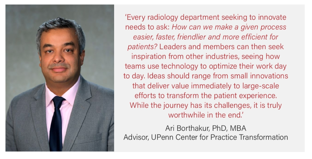
Submitted by Ari Borthakur, PhD, advisor at Penn Medicine’s Center for Practice Transformation
