Scientists look to functional MR brain imaging to understand America’s political divide
As America looks to heal its political division following this bruising election cycle, scientists are turning to functional MR brain imaging for answers.
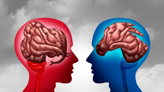
Experts from three prominent institutions recently scanned the minds of more than three dozen conservative- and liberal-leaning individuals, looking for insights. While viewing videos related to hot-button immigration issues, researchers discovered that the two sides’ brains respond differently to the same stimuli.
Divergences were even more noticeable when videos contained words that commonly crop up in campaign ads, according to their analysis, published in the Proceedings of the National Academy of Sciences.
“Our study suggests that there is a neural basis to partisan biases, and some language especially drives polarization,” lead author Yuan Chang Leong, PhD, a postdoctoral scholar in cognitive neuroscience at UC Berkeley, said in a statement. “In particular, the greatest differences in neural activity across ideology occurred when people heard messages that highlight threat, morality and emotions.”
For their analysis, researchers from Berkley, Stanford University and John Hopkins recruited 38 young and middle-aged individuals with similar socioeconomic and educational backgrounds and strong views on immigration. They scanned participants’ brains using fMRI while they viewed two dozen videos highlighting liberal and conservative positions on the topic. And after each, viewers rated their level of agreement with the videos on a scale of 1-5.
Leong et al. noted that both sides showed a high shared response in the auditory and visual cortices. But neural responses diverged along partisan lines in the dorsomedial prefrontal cortex, where semantic information, or word meanings, are processed, the team noted. In particular, words related to “risk and threat” and “morality and emotion” produced greater polarization in each participant’s neural response.
For further research, Leon and colleagues are looking to use neuroimaging to craft more precise models of how individuals interpret political content. They hope this may inform interventions that narrow the political divide between the two parties.
“We know that partisans respond differently to the same information. So in that sense, it's not surprising to find that their brains respond differently as well," Leong told Medscape. “If our goal is to reduce polarization and change minds, we need to think carefully about how we frame and structure political information—for example, by framing messages to appeal to the core values of the respective voter," he added later.
Read more about their investigation in PNAS here.

