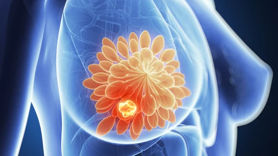
Simple, readily available MRI measurement could reduce breast biopsies by one-third
A simple, readily available MRI measurement could potentially reduce breast biopsies by almost 33%, according to a new study out of Austria.
MRI is an oft used means of detecting and classifying tumors, but it can sometimes result in false positives leading to unnecessary biopsies, excess costs and overtreatment. Scientists with the University of Vienna, however, have validated a noninvasive imaging biomarker they say can reduce biopsies after MRI.
The practice change would only take about three minutes, can be incorporated into standard short-MRI scans, and uses infrastructure that exists in most radiology practices. Diffusion-weighted imaging is already established in stroke care and is becoming increasingly popular in cancer diagnostics.
“It can be used anywhere, since it is largely independent of the equipment, the experience of the radiologist, the measuring time and the measuring technique,” principal investigator Pascal Baltzer, with the Department of Biomedical Imaging and Image-guided Therapy at MedUni Vienna, said March 1. “Our finding can be used immediately for diagnostic purposes. It does not require a special center—every registered radiology department would be able to use it straightaway.”
Contrast-enhanced magnetic resonance imaging helps to measure blood circulation in tissue, but radiologists sometimes have difficulty pinpointing whether nodes are malignant or just well-supplied with blood. Coupling this with diffusion-weighted imaging, however, aids in mapping the motion of water molecules in investigate structures, Baltzer and colleagues noted, and allows providers to measure it objectively using the apparent diffusion coefficient.
“DWI enables us to characterize lesions much better because hydrogen molecules ‘dance’ faster in healthy tissue than they do in malignant tumors,” said lead author Paola Clauser.
University of Vienna scientists tested the measure in a retrospective study using data from five centers and 657 female patients. They found the apparent diffusion coefficient logged a sensitivity rate of 96.6%, with the potential reduction of unnecessary biopsies at 32.6%.
You can read more about their results in Clinical Cancer Research here.

