MRI after button battery removal: Consensus on next steps lacking among referrers
Consensus is lacking among referring physicians on how imaging findings should guide next steps following the removal of an ingested button battery, according to survey data published Wednesday.
Medical societies recommend gathering cross-sectional imaging of pediatric patients in these instances. However, there are no published guidelines on how such scans should inform clinical management, experts wrote in Clinical Imaging.
Seattle Children’s Hospital experts are calling for further studies to help fill this void of information.
“The management of button battery ingestion patients poses a unique challenge due to the risk of life-threatening complications weeks after endoscopic foreign body removal,” Teresa Chapman, MD, a University of Washington professor and pediatric radiologist at Seattle Children’s, and colleagues wrote March 23. “Further multicenter research is needed to inform development of updated clinical management tools and to determine if MRI can effectively predict vascular emergencies and prevent catastrophic patient outcomes.”
Chapman and a working group of providers surveyed 42 pediatric subspecialists in their organization for the study. Half responded (22), including four otolaryngologists, nine general surgeons and nine gastroenterologists. Answers revealed no agreement among docs on how battery location might impact their decisions. About 68% said acute necrosis of the esophagus was cause for intensive care unit admission, while 54% said mucosal necrosis was a reason to delay the initiation of oral feeds. Another 59% felt this was an indication for surgical consultation, and 54% said that neither mucosal necrosis nor battery location dictated the timing of hospital discharge.
Providers were also polled on MRI and CT angiography findings of interest. All said evidence of tracheoesophageal fistula would prompt ICU admission. Another 95% said diagnosis of this abnormal connection between the two tubes, causing foods and fluids to aspirate into the lungs, would delay transition to oral feeds. About 68% considered it cause for surgery, while most said swelling abutting a major artery (86%) or vein (77%) would result in an ICU admission.
“Our working group's recommendation for our own institution is that MRI be performed one week following button battery removal to evaluate for mediastinal fluid collection or spine infection,” Chapman et al. wrote. “We currently reserve repeat MRI for cases in which the patient does not respond to supportive therapy and antibiotics as expected. Fluoroscopic esophagram and timing of resumption of oral feeds is at the discretion of the care team depending on extent of esophageal injury.”
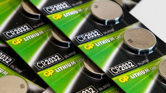
The CDC has reported a more than twofold increase in injury from these batteries among children under 13 and sevenfold increase in severe complications or death. Experts have attributed the trend to production of larger, high-voltage lithium cells, the study’s authors noted.

