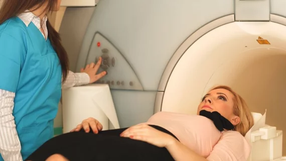
Deep learning expedites normal findings on ultrafast breast screenings
AI can safely and accurately identify healthy breast tissue on ultrafast breast MRI, negating the need for a radiologist’s closer look and, in the process, lowering cancer screening costs and widening patient access to breast MRI.
So conclude researchers at the University of Groningen Medical Center in the Netherlands whose report is running in European Radiology [1].
Xueping Jing, Peter M.A. van Ooijen and colleagues trained a deep learning model on 837 breast MRI scans from 438 women that were labeled at final interpretation as either normal (without suspicious lesions) or abnormal (with suspicious lesions).
Testing the model as tuned to a default probability threshold on an independent image set of 178 scans from 149 patients, the team found it brought back an AUC of 0.81. When they applied a high-sensitivity threshold, the researchers found their algorithm yielded a sensitivity of 98%, a negative predictive value of 98%, a workload reduction of 15.7% and a scan time reduction of 16.6%.
Further, these process improvements came at the modest clinical cost of 1 of 55 scans getting incorrectly read by the AI as negative.
“Although a conservative strategy was adopted, there were still false negative predictions,” the authors comment in their discussion. However, all the missed lesions were smaller than 10 mm, they point out, and “the relatively small size may be the main reason that the deep learning model did not detect them.”
In addition, 134 of 195 false positive predictions were BI-RADS 2, and 113 were assessed within heterogeneous and extreme fibroglandular tissue.
“This finding indicates that proper handling of dense and BI-RADS 2 breasts may be the key to reducing false positives in the future,” the authors write.
More from the discussion:
[T]he challenge of using AI in triaging is the same: A lower threshold is safer but less efficient, and the trade-off between the risk of missing breast cancer and the reduction of workload makes the threshold difficult to determine. … [T]he classification of ultrafast breast MRI examinations with a deep learning model in the workflow may be a promising method to improve the efficiency and accessibility of breast MRI screening.”
The journal has posted the study in full for free.
More Coverage of Breast MRI:
Abbreviated breast MRI deemed an attractive screening option—sometimes
Screening breast MRI results in more downstream healthcare costs than mammography alone
Follow-up imaging for incidental liver lesions on breast MRI rarely necessary
Simple switches help hospital reduce excess patient visits, biopsy wait times after breast MRI
Contrast-enhanced mammography as effective as MRI at evaluating newly diagnosed breast cancer
Reference:
- Xueping Jing, Peter M.A. van Ooijen et al., “Using deep learning to safely exclude lesions with only ultrafast breast MRI to shorten acquisition and reading time.” European Radiology, May 26, 2022. DOI: https://doi.org/10.1007/s00330-022-08863-8

