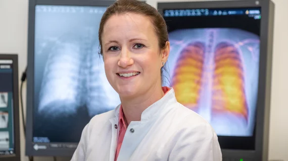
Dark field chest X-rays can improve diagnosis of COVID-related lung disease
A novel X-ray technology known as dark-field chest X-ray can offer key information about COVID-related lung disease, according to researchers at the Technical University of Munich (TUM) in Germany.
Using small-angle X-ray scattering, dark-field chest X-rays help physicians visualize the lung tissue’s microstructure, explains Franz Pfeiffer, a professor of biomedical physics and director of the Munich Institute at TUM.
“During our X-ray examination, we take conventional X-ray images and dark-field images simultaneously. This gives us additional information about the affected lung tissue quickly and easily,” Pfeiffer tells the school’s news operation.
With a radiation dose fifty times smaller than computed tomography imaging, dark-field chest X-rays could potentially serve a key purpose as a lower-radiation alternative to CT, especially in cases requiring ongoing observation that would otherwise result in heavier radiation exposure.
“Consequently, this method is promising for application scenarios that require repeated examinations over extended periods of time—for example, when investigating the progression of long COVID. This approach might serve as an alternative to computed tomography for imaging lung tissue over prolonged observation periods," Pfeiffer says.
Beyond COVID-19, dark-field chest X-rays may also be useful for evaluating other instances of lung disease unrelated to the pandemic. Inflamed lung tissue will produce a weaker dark-field signal, allowing physicians to hone in on potential lung damage where the tissue appears darker in the image.
To quantitatively evaluate the darker signals while adjusting to account for variations between individuals, the research team produced a prototype of an “optimized” X-ray machine. Daniela Pfeiffer, professor of radiology and medical director of the study, describes the potential to further assist physicians with evaluation in the future through developing AI options.
“Once we have sufficient dark-field X-ray data available, we intend to use artificial intelligence to support the evaluation process,” she says. “For conventional images, we have already carried out a pilot project for AI evaluation of our X-ray images."
