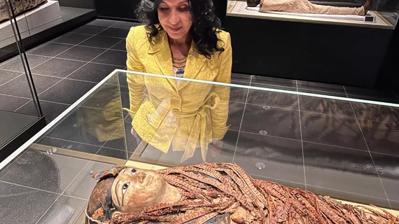
Paleo-radiologist: ‘I saw the king’s face for the first time’
Thanks to a radiologist equipped with CT and 3D image reconstruction, Pharaoh Amenhotep I is finally getting a proper clinical workup.
The monarch reigned over an Egyptian dynasty in the early 1500s BC and was discovered among several dozen other mummies in the 1800s.
The radiologist, Sahar Saleem, MD, of Cairo University, a renowned Egyptologist, says the king died at around 35 years old while in good health (which may suggest regicide).
She also says the scanning revealed Amenhotep I is adorned with 30 amulets and wearing a belt made with 34 golden beads.
This is an important discovery, Saleem says, because the jewels and accessories would seem to refute earlier theories that subsequent dynastic generations cared for rather than robbed their past rulers.
Middle East Eye reported on the development Jan. 12.
Dr. Saleem says she believes her CT findings represent the best archeological discovery in the world, not just in Egyptian antiquities.
Archaeology aficionados are especially excited about the discovery, she suggests, because they are now “able to see the face of King Amenhotep I, the son of King Ahmose, conqueror of the Hyksos, after 3,000 years.”
She’s referring to the face revealed by a 3D image reconstructed from images of the skull and facial bones.
“I examined the mummy through CT scans without peeling away the scrolls or causing any damage to the organic remains,” she tells Middle East Eye reporter Salwa Samir. “I saw the king’s face for the first time.”
The beauty of CT as applied to mummies is its relative gentleness, Dr. Saleem underscores with evident enthusiasm.
In the past she used 3D printing to build shoulders-and-up busts of historically important mummies, including famous King Tut.
As for Pharaoh Amenhotep I, his mummy has been a source of speculation and guessing ever since its initial discovery in 1881. That’s largely because respectful historians have been reluctant to risk damaging it while conducting examinations, reporter Samir points out.
Now, thanks to CT and 3D, Egyptology has a paleo-radiologist dedicated to caring for its beloved ancient “patients.”
“On my first day in the hospital [in 2004], they brought an Egyptian mummy for a CT scan,” Dr. Saleem reflects. “I thought immediately that I can benefit my own civilization through specializing in this field. [Now] I have the ability to understand my civilization and be its guardian.”

