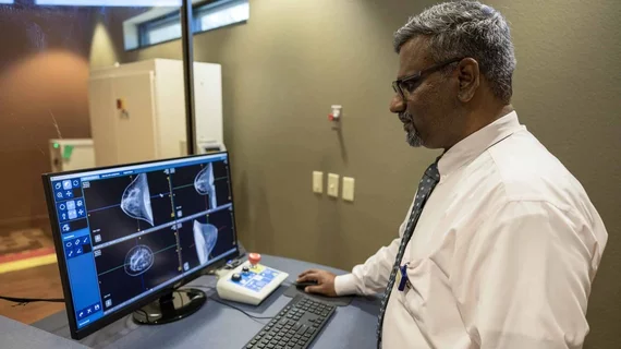
Radiology researchers score $3M NIH grant to develop compression-free breast CT system
Radiology researchers with the University of Arizona Health Sciences have scored a $3.3 million grant from the National Institutes of Health to develop a new compression-free breast imaging system.
They’ll use the funds to continue testing their computed tomography technology, which eliminates tissue overlap and instead uses CT to create high-resolution images.
The system includes a table with a hole in the middle. Women lie prone with one breast through the opening, and the tube spins around 360 degrees, noted Srinivasan Vedantham, PhD, a professor in the U of A College of Medicine–Tucson’s Department of Medical Imaging. The system makes no contact with the breast and takes about 10 seconds to image each side of the chest.
“The advantage of breast CT is we have a whole 3D image. It would have better sensitivity in detecting breast cancer, particularly for women with dense breasts,” Vedantham said in an Oct. 21 announcement from the university. “Our goal is to enhance breast cancer screening technology to improve early detection and outcomes for patients.”
Vedantham and colleagues will use the government grant to further refine their noncompression CT scanner prototype. They’ve already used the system on 92 women in collaboration with the U of A Cancer Center. The university hopes to recruit an additional 600 volunteers to test the CT system and compare it against 3D mammography.
University of Arizona Health Sciences experts previously earned a $3.6 million grant from the NIH to develop the technology in 2019.

