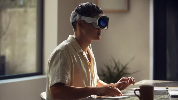
Next-generation VR headsets could improve ergonomics, radiologist reading experience
Next-generation VR headsets are showing promising in improving the radiologist experience while interpreting medical images.
Researchers with the University of California, San Diego, recently tested this new virtual reality technology, sharing their work Monday. They utilized the Visage Ease VP software on an Apple Vision Pro headset, comparing diagnostic performance for detecting diverticulitis on CT.
Radiologists’ results were similar between using the augmented-reality headset and Visage 7 on a desktop diagnostic monitor, experts wrote in the Journal of Imaging Informatics in Medicine [1]. However, ease of use, experience and preference varied among physicians.
“The headset offers new opportunities for 3D visualization through its immersive display and new possibilities for ergonomics through the eye and hand tracking of its user interface,” Paul Murphy, MD, PhD, an associate clinical professor of radiology at UCSD, and co-authors wrote Nov. 4. “For these reasons, next-generation VR/AR headsets are promising for many applications throughout radiology.”
The pilot study retrospectively included 110 patients who underwent abdomen/pelvis CT scans for which the report mentioned the presence or absence of the gastrointestinal disease. Scans were matched by time and other factors for a total of 55 cases with diverticulitis and 55 controls without. Six radiologists practiced using the headset and app on 10 scans. They then each scored 50 unknown images on a desktop and 50 more via VR, using a six-point scale for diverticulitis—with 1 indicating no disease and 6 the opposite.
Afterward, radiologists completed a survey on the ease of use for the headset and viewer app. The area under the curve (and 95% confidence interval) for diverticulitis was 0.93 with the headset and 0.94 using a desktop. Median time per case was about 57 seconds for the headset and 31 for the desktop. Meanwhile, average survey scores (on a 5-point scale) ranged from 3.3 to 5 for ease of use, 3 to 4.7 for experience, and 2.2 to 3.3 for preference.
Each reader utilized the “guest user mode” on the headset to help calibrate the eye- and hand-tracking interface. Readers who required them wore contact lenses and were oriented to the device by performing several tasks to view scans and 3D constructions. Radiologists assessed the visibility of 5% and 95% luminance boxes and of 1-and 2-pixel line pairs of an 800 x 600-pixel TG18 test image, chosen due to their similarity with the CT matrix size of 512 x 512, rather than the resolution of the headset display. Readers also sat during most of the study but were required to rotate their heads to view different parts of the app, the authors noted.
“Given the importance of CT within radiology, it is important to address whether its highest-resolution features can be visualized on imaging displays such as those of next-generation VR/AR headsets,” Murphy and colleagues added. “This pilot study found no significant difference in the pooled diagnostic performance for diverticulitis between radiologists using a headset versus a desktop, and radiologists reported an overall good user experience with the headset. These results suggest that the display of this headset may suffice for visualization of such features.”

