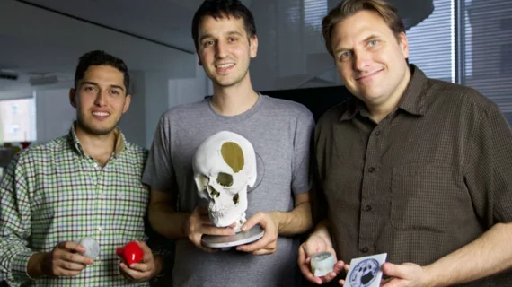
Novel 3D-printing technique generates full anatomical models from MRI, CT scans
A 3D-printing technique originated at Harvard University allows clinicians to produce highly detailed models of human anatomy in less than an hour—for a fraction of the cost and labor needed for a lower quality product, researchers reported in 3D Printing and Additive Manufacturing this month.
The method was born from 26-year-old grad student Steven Keating’s wish to physically hold and visualize his brain in real-time after he had a baseball-sized tumor removed from his brain, the authors said. Keating, who now holds a doctorate from MIT, said he wanted to better understand diagnosis and treatment options after his cancer scare but was unable to produce the results he wanted with available 3D-printing technologies.
Keating collaborated with a team of Harvard scientists headed by James Weaver, PhD, a senior research scientist at the Wyss Institute, to create a technique that translates MRI and CT images into 3D models of “unprecedented detail.”
“Our approach not only allows for high levels of detail to be preserved and printed into medical models, but it also saves a tremendous amount of time and money,” Weaver said in a release from Harvard. “Manually segmenting a CT scan of a healthy human foot, with all its internal bone structure, bone marrow, tendons, muscles, soft tissue and skin, for example, can take more than 30 hours, even by a trained professional. We were able to do it in less than an hour.”
The team started with conventional MRI and CT scans—including Keating’s—that they fed into 3D printers. Since advanced imaging builds images through a series of high-resolution slices, they said, and 3D printers build models in a layer-by-layer process, the two technologies married well. Development wasn’t all easy, though—Weaver and co-authors said that because MRI and CT both produce such highly detailed images, it was initially difficult for the printer to distinguish which areas of the scan to actually print.
The researchers used bitmap-based printing, an alternative to the arduous process of segmentation, to achieve a better product. Dithered bitmaps are able to simplify an image’s pixels from grayscale to black and white pixels, they wrote, so the 3D printer would be able to distinguish its area of interest from surrounding tissue.
Soon, Keating was holding a detailed model of his brain—something Weaver said he hopes isn’t too far off for others.
“I imagine that sometime within the next five years, the day could come when any patient that goes into a doctor’s office for a routine or non-routine CT or MRI scan will be able to get a 3D-printed model of their patient-specific data within a few days,” he said.
Keating said in the release he routinely uses the new technique to keep up with his health and check on his skull’s recovery after the tumor removal.
“The ability to understand what’s happening inside of you, to actually hold it in your hands and see the effects of treatment, is incredibly empowering,” he said.

