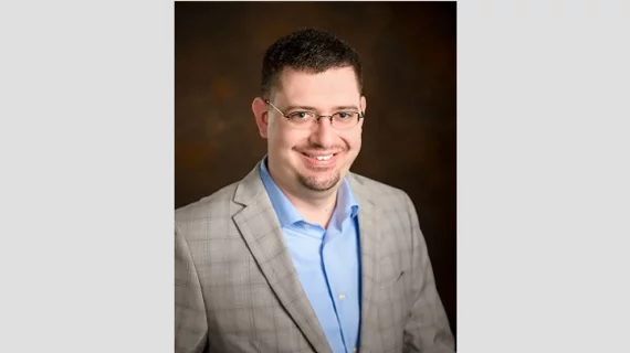
Q&A: The new era of AI in medical imaging, and what it means for patients
Artificial intelligence (AI) has been one of the most hyped advancements in radiology for many years now, sparking vigorous debates about its potential impact on patient care. But early on, discussions about AI were more theoretical than factual; it was simply too early to know what the future may bring.
The times, however, they are a-changin’. Brian Baker, director of software engineering at vRad, a MEDNAX Company, has been tracking automation for his entire career and leads many of vRad’s AI efforts. He says 2019 is an exciting new time for the relationship between AI and radiology, a time when the world is finally starting to see just how impactful these technologies can be on a day-to-day basis.
Baker spoke with us about AI’s influence in today’s healthcare marketplace, how vRad and MEDNAX are working with these evolving technologies and more. The full conversation is below:
What excites you the most about AI in medical imaging right now?
Brian Baker: The most exciting aspect of AI in medical imaging is that we are starting to see a real impact on patients. We are beyond theory and academic papers and are implementing substantial, results-driven solutions.
At MEDNAX, the most meaningful advances are ones that tangibly help patients—prioritizing worklists based on the probable presence of a condition, for example. We are also using AI to help make radiologists more efficient—pulling data from worksheets, determining orientation of images, and so on—but our passion is patient-centric. MEDNAX has moved past writing academic papers with lab data; we have validated our AI models and are using them in real time to improve patient care.
What are some examples of what MEDNAX and vRad are doing with AI?
Right now, MEDNAX has models that score studies for the probability of various pathologies: acute intracranial hemorrhages, pulmonary embolisms, aortic dissections and pneumothorax. These results can be used to adjust worklist prioritization, ensuring that patients with a high probability of positive, critical conditions are getting treated as quickly as possible. We are able to positively impact 18 patients per day on average, getting their studies read up to 15 minutes faster. The AI models currently in production have also identified possible critical findings on around 20 studies per month that were originally ordered as non-emergent ‒ allowing our system to escalate the orders, getting the studies to the radiologists potentially 24 hours sooner. For patients with critical conditions, those minutes and hours are crucial.
What do the radiologists at MEDNAX think about AI?
While I certainly can’t speak for all radiologists at MEDNAX, I hear a few common threads. I do know there are occasional concerns about AI replacing radiologists, but I am personally confident it will serve to augment rather than replace radiologists.
More commonly, I hear questions on how AI will change radiology and how radiologists can get involved. AI is moving into the workflow at MEDNAX and many radiologists are eager to participate in a meaningful way. This is fortunate, because the medical imaging AI field is in acute need of radiology expertise. As you probably know, the single most important factor in success in AI is well-annotated data. We need radiologists’ help to annotate a lot of images.
We also take close direction from MEDNAX radiologists on which AI solutions to build and which should be a top priority. My team is fortunate to have a number of radiologists patient enough to pair closely with our data scientists to define use cases, annotate images, explain diseases and conditions, and provide guidance as AI models are developed. Clinical expertise is a cornerstone of our processes at MEDNAX; radiologists are involved throughout the development and validation processes, making key decisions to ensure our solutions have high clinical value.
Can you give us an idea of how MEDNAX goes about validating the models you describe?
One of the core tenants of the AI efforts at MEDNAX is to ensure that the models work with our extremely heterogeneous data set. We call this “validation in the wild.” With more than 2,000 facilities across the United States, we get an extremely diverse set of patients and very different combinations of factors within the DICOM tags and images. Each model at MEDNAX is put through a set of stages we call “the funnel” and each stage requires more rigorous testing.
We also use natural language processing (NLP) to “measure” the model and determine specificity and sensitivity. While we do measurements in the lab on large data sets, we must know how a model performs operationally with hundreds of thousands of studies. To do this, we utilize NLP to “read” the reports and then use that data as a comparison against the model.
There are some important nuances. The NLP measurements are specific to the pathology and use cases. For example, let’s examine intracranial hemorrhages (ICH). For our AI model that scores the probability of a study being positive for acute ICH, our NLP measurement is designed to find studies that have acute, non-stable ICH reported by the radiologist. This means that any chronic (non-acute) or stable (unchanging or shrinking such as a follow up study) ICH will be marked as a negative by the NLP measurement. This is important because the NLP is measuring for the use case of worklist prioritization and this use case is specific to acute, unstable hemorrhages.
When we measure our own AI model or a partner’s model, that model was most likely validated in the lab with different clinical criteria—perhaps for ICH, the model was trained to look for all bleeds, regardless of acuity or stability. MEDNAX is careful to measure a model against the use case in mind, with special care to the clinical variations in usage.
For more on the MEDNAX AI program
vRad Presents at SIIM Meeting on Artificial Intelligence Model to Assess Probability of Aortic Dissection – June 27, 2019
Beyond the hype: How practical AI is enhancing radiology
Imad B. Nijim, Chief Information Officer, MEDNAX Radiology Solutions

