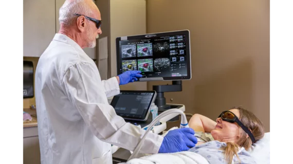
Breast specialists have seen the future. It’s opto-acoustic imaging.
Few radiology subspecialties can match breast imaging at adopting new imaging modalities to improve patient care. Consider: The past 10 to 15 years have seen the rise of digital breast tomosynthesis, automated breast ultrasound, breast MRI (with and without diffusion weighting), low-dose positron emission mammography and contrast-enhanced dual-energy digital mammography. That’s just to name a few.
What these technologies have in common is the profession’s interest in screening as many patients as possible for cancer without sending anyone for unnecessary biopsies or unwarranted follow-up exams.
To be sure, this aim remains largely aspirational. Recent research has shown almost 50% of patients recalled from screening for a possible abnormality receive two follow-up diagnostic exams before arriving at a definitive diagnosis. Close to 20% have three follow-ups and around 10% have four.
As for biopsies, for every 10 women requiring diagnostic evaluation after screening, one will undergo the stressful and uncomfortable procedure. Despite the trouble and worry, masses in these women turn out to be benign in 75% of cases.
It doesn’t have to be this way. Numerous experts agree that opto-acoustic ultrasound imaging—or “OA/US,” as it’s called for short—is the most revolutionary of all the newer breast-imaging modalities.

“Opto-acoustic breast ultrasound gives us the opportunity to make a much better decision about marginal lesions than we could ever get before.”
Michael Linver, MD, Clinical Professor of Radiology, University of New Mexico School of Medicine and Adjunct Professor of Radiology, George Washington University School of Medicine
When used as the first diagnostic exam following an abnormal screening mammogram that demonstrates a non-calcified mass of uncertain pathology, OA/US has been proven capable of reducing false positives—and thus unneeded biopsies—by some 75%.
The above statistics come from objective, peer-reviewed research.1,2 The subjective perspective is from several radiologists who are familiar with opto-acoustic imaging and spoke about it with Radiology Business.
“Opto-acoustic breast ultrasound gives us the opportunity to make a much better decision about marginal lesions than we could ever get before,” says Michael Linver, MD, a breast imaging specialist who has been practicing, teaching and conducting clinical research since the 1970s.
“Up to now, regardless of imaging modality, many lesions imaged diagnostically were placed in the BI-RADS 3 category,” adds Linver, clinical professor of radiology at the University of New Mexico School of Medicine and adjunct professor of radiology at George Washington University School of Medicine. “Often this was because the radiologists thought the lesions were benign but weren’t sure, so they made patients come back in six months to check again.”
Anatomy and function
Here’s how it works. Opto-acoustic imaging takes the central element of standard ultrasound—reflected sound waves, aka “acoustics”—and adds noninvasive laser light (hence “opto,” short for optical). The combination allows the radiologist to interpret noninvasive imaging findings along two complementary tracks.
The high-resolution ultrasound shows static anatomic features such as lesion size, location and shape. The laser optics reveal functional properties that help to clearly identify or rule out malignancies.
Most important among the optically exposable properties are angiogenesis and hypoxia. If angiogenesis is present, the mass is growing new blood vessels to supply itself with oxygen and nutrients. Similarly, positive findings for hypoxia mean the lesion is overusing its blood supply and impacting both structural and functional characteristics in and around the tumor.
Other indicators are used, but if both angiogenesis and hypoxia are at work, the mass is probably a cancer.
OA/US technology has been around since the first decade of the 2000s, but, as Linver points out, it has “matured dramatically” over the last five years or so.
“We now have an opportunity to take a large segment of that ‘BI-RADS 3’ group and characterize their masses, quickly and with great confidence, as either positive or negative,” Linver says. “This is a huge plus for patients, who get very upset when called back for more examinations that ultimately turn out to have been needless.”
“In my opinion,” Linver offers, “avoiding those unnecessary callbacks is really the greatest gift opto-acoustic imaging offers to those who use it.”
Cost-effective choice
The technology may be in the best interest of not only patients and radiologists but also U.S. healthcare as a whole. Earlier this year, radiology researchers at the University of Texas published a decision-tree modeling study examining the value of supplemental opto-acoustic imaging for breast masses that had been flagged in screening. Lead author B. Bersu Ozcan, MD, and colleagues found OA/US to be more cost-effective than standard ultrasound for distinguishing benign from malignant masses.
Conducting a probabilistic sensitivity analysis as part of the study, they found opto-acoustic imaging the better strategy in 98.69% of 10,000 iterations.
OA/US, the study concluded, “can reduce costs while improving patients’ quality of life, primarily by reducing false-positive results with consequent benign biopsies.”3

“Opto-acoustic imaging is fast, simple and can dramatically improve diagnostics.”
Bersu Ozcan, MD, Department of Radiology, UT Southwestern Medical Center
Ozcan and co-author Basak Dogan, MD, expounded on their findings for Radiology Business.
“One of the best things about opto-acoustic imaging is that we are not injecting any contrast,” Ozcan says. There’s no ionizing radiation, either, and yet “we are getting additional real-time functional information in addition to conventional ultrasound images.”
Ozcan underscores that the cost-effectiveness study only compared OA/US with conventional gray-scale ultrasound, specifically in a diagnostic setting. Further, it was a modeling study using previously published data. As a result, the researchers could not incorporate all potential downstream expense and utility, like cost related to technologist training. Nevertheless, she says, “we can say that OA/US improves patients’ quality of life and overall lowers their costs.”
Asked for her own judgment—based on not just the study at hand but also on her full professional experience with the technology—Ozcan replies:
“Opto-acoustic imaging is fast, simple and can dramatically improve diagnostics.”

The real innovation of opto-acoustic imaging “lies in its ability to eliminate the need for contrast, reduce the necessity for biopsies through enhancing the diagnostic value of ultrasound and, in some cases, even avoid surgery—all while still providing a precise understanding of the disease.”
Basak Dogan, MD, Director of Breast Imaging Research, UT Southwestern Medical Center
A technology that ‘could be making a huge difference’ right now
Dogan concurs. “Each cancer is unique,” she explains. “Treatment must be tailored to the individual case. There’s no ‘one-size-fits-all’ approach.”
The real innovation of opto-acoustic imaging, she adds, “lies in its ability to eliminate the need for contrast, reduce the necessity for biopsies through enhancing the diagnostic value of ultrasound and, in some cases, even avoid surgery—all while still providing a precise understanding of the disease.”
This includes assessing lymph node involvement, she notes, which is a critical indicator for predicting the potential spread of the cancer.
“If we can unlock how a cancer will behave based solely with imaging, we can better determine which patients might benefit from surgical lymph node evaluation,” Dogan says. “When you combine that with the ability to track chemotherapy response using OA/US, the value of this technology becomes clear.”
For Linver, the biggest concern is that not as many breast imagers and referrers are hearing about opto-acoustic imaging as ought to.
“I wish we could get the technology in front of more professionals so they can appreciate the difference between OA/US and standard ultrasound,” Linver says. “Opto-acoustic ultrasound imaging could be making a huge difference for breast specialists and their patients right now. This is a technology that I really believe in.”
References:
- Vlahiotis, A., Griffin, B., Stavros, A.T., & Margolis, J. (2018). Analysis of utilization patterns and associated costs of the breast imaging and diagnostic procedures after screening mammography. ClinicoEconomics and Outcomes Research: CEOR, 10, 157–167. https://doi.org/10.2147/CEOR.S150260
- Lang, E.V., Berbaum, K.S., & Lutgendorf, S.K. (2009). Large-core breast biopsy: abnormal salivary cortisol profiles associated with uncertainty of diagnosis. Radiology, 250 (3), 631–637. https://doi.org/10.1148/radiol.2503081087
- Ozcan, B.B., Xi Y., Dogan, B.E. (2023). Supplemental optoacoustic imaging of breast masses: a cost-effectiveness analysis. Acad Radiol 31(1):121–130. https://doi.org/10.1016/j.acra.2023.08.042

