
Beyond AI: How 3D Surgical Intelligence Is Expanding Radiology’s Clinical Impact
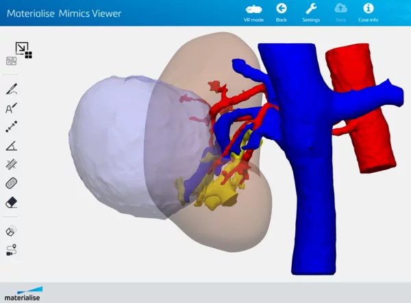
Exploring how 3D surface modeling workflows are expanding radiology’s role in multidisciplinary surgical care.
3D surface modeling and virtual surgical planning are expanding radiology’s contribution to multidisciplinary teams. Together, AI-driven interpretation and 3D-enabled clinical collaboration represent two distinct, complementary forces shaping the next decade of radiology.
Beyond Automation: Delivering Imaging Insights Surgeons Can Interact with
Surgical preparation often requires more than the identification of pathology. Surgeons frequently need a detailed understanding of patient-specific anatomy to see the relationship between vessels, tumors, bones, and soft tissue structures that can be explored interactively before intervention.
This is where 3D surface modeling plays a critical role. As 3D technologies mature, the distinction between volume rendering and surface modeling has become increasingly relevant for radiology departments evaluating tools for surgical planning.
Traditional 3D volume rendering software within PACS is a representation derived from 2D imaging data that displays voxel intensity throughout a volume, ideal for routine diagnostics. In contrast, 3D surface modeling software uses segmentation to extract specific anatomical structures and convert them into discrete, accurate 3D geometric models.
These models can be rotated independently, and individual structures can be shown or hidden to reveal underlying anatomy, providing greater clarity for assessing spatial relationships than volume-based approaches allow.1 Surgical teams can also evaluate procedural approaches virtually before entering the operating room, helping align imaging findings with surgical strategy.
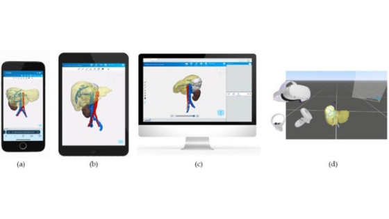
Radiologists who integrate structured 3D surface modeling workflows into their practice are not replacing diagnostic interpretation but extending it. By translating imaging data into interactive, patient-specific models, they provide insights that help surgical teams plan complex procedures with greater confidence.
Diverse published evidence demonstrates the clinical impact of this approach: studies document measurable reductions in operating room time, fewer intraoperative complications, and improved surgical predictability when surgical teams plan with patient-specific 3D surface models.2-4
Multidisciplinary decision-making
At leading institutions, 3D planning with surface modeling is increasingly embedded into routine workflows for complex cases such as organ-sparing surgeries, orthopedic reconstructions, tumor resections, and breast reconstruction procedures. Published experiences, including work from institutions such as Mayo Clinic and other academic surgical centers, document how patient-specific 3D surface models and 3D planning improve preoperative understanding and coordination across teams.
In these settings, radiology-produced 3D surface models become a shared reference point during case discussions. Rather than relying solely on descriptive reports, surgeons and radiologists interact with the same digital anatomy model. This shared visual language enhances communication, clarifies anatomical variability, and supports more informed decision-making.
Digital planning vs. physical printing: lowering the barrier to entry
When 3D technology is discussed, many departments immediately think of 3D printing labs and capital-intensive infrastructure. In reality, much of the clinical value lies in digital planning workflows rather than the manufacturing of physical models.
3D surface modeling provides detailed anatomical clarity without the time, cost, or logistics of printing. Digital models can be updated instantly, manipulated dynamically, and shared securely across teams. Extended reality platforms further extend visualization capabilities without requiring physical production.
Advanced 3D planning platforms, such as Materialise Mimics, integrates directly into existing imaging workflows. Radiologists can generate interactive models from their workstations and share them securely with surgical colleagues. For many departments, this means adoption does not require a dedicated lab but rather structured workflows, training, and defined use cases.
Reimbursement Signals Growing Recognition
A historic milestone in the United States underscores the maturation of 3D surface medical workflows. In September 2025, the AMA CPT Editorial Panel approved the first formal coding recognition for 3D surface modeling, digital surgical planning, and extended reality (XR) applications.
Effective July 1, 2026, six new Category III CPT codes specifically support software-based 3D planning workflows, enabling radiology departments to implement software-based surgical planning without requiring 3D printing infrastructure. These codes cover 3D surface modeling (1030T, +1031T), digital surgical simulation including VR/XR applications (1032T, +1033T), and computational modeling (1034T, +1035T), with time-based add-on codes for extended work.
The codes are temporary designations that establish a clear reimbursement pathway, with monetary values currently under discussion. These efforts, coordinated by leaders such as Dr. Frank Rybicki and supported by the RSNA 3D Printing Special Interest Group, aim to build the clinical evidence needed to transition these codes to permanent Category I status within five years. Radiology departments implementing 3D workflows can contribute to this effort by participating in clinical registries.
Implementation: Starting Focused, Scaling Strategically
Successful 3D adoption rarely starts as a large, enterprise-wide initiative. Most programs begin with targeted pilot cases in specialties where anatomical complexity makes the benefit immediately visible, such as orthopedics, oncology, cardiology, and urology.
Radiologists and technologists with strong anatomical knowledge can be trained efficiently in surface modeling workflows. By positioning 3D surface modeling as a radiology-owned service, departments maintain quality control, streamline collaboration, and avoid fragmentation across engineering or external units.
The result is a scalable model that grows alongside clinical demand.
AI and 3D: Complementary Forces Shaping Radiology’s Future
AI will continue to enhance image interpretation and operational efficiency. At the same time, 3D surface modeling expands the role of imaging data in procedural care. These trends are not in conflict; they are complementary.
As automation enables faster, more consistent readings, radiologists have new opportunities to expand their impact through advanced visualization and multidisciplinary collaboration. Together, AI-driven analysis and 3D-enabled planning define a future in which radiology is both technologically advanced and deeply integrated into patient-specific procedural care.
For departments evaluating their strategic direction, the question is no longer whether 3D planning is feasible. With mature software platforms, established workflows at hundreds of institutions, and emerging reimbursement pathways, the infrastructure is in place. The opportunity lies in determining how imaging insights can best support the evolving needs of surgical teams and in radiology's continued leadership at the intersection of diagnosis and clinical application.
Discover how 3D surface modeling can support your department’s strategic growth.
Visit: www.materialise.com/medical
References
- Valls-Esteve A, Adell-Gómez N, Pasten A, Barber I, Munuera J, Krauel L. Exploring the Potential of Three-Dimensional Imaging, Printing, and Modeling in Pediatric Surgical Oncology: A New Era of Precision Surgery. Children. 2023;10(5):832. doi:10.3390/children10050832
- Stepanenko A, Perez LM, Ferre JC, et al. 3D Virtual modelling, 3D printing and extended reality for planning of implant procedure of short-term and long-term mechanical circulatory support devices and heart transplantation. Front Cardiovasc Med. 2023;10:1191705. doi:10.3389/fcvm.2023.1191705
- Sanchez-Garcia J, Lopez-Verdugo F, Shorti R, et al. Three-dimensional Liver Model Application for Liver Transplantation. Transplantation. 2024;108(2):464-472. doi:10.1097/TP.0000000000004730
- Wake N, Rosenkrantz AB, Huang WC, et al. A workflow to generate patient-specific three-dimensional augmented reality models from medical imaging data and example applications in urologic oncology. 3D Print Med. 2021;7:34. doi:10.1186/s41205-021-00125-5