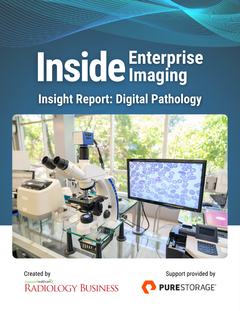
Pathology Is Going Digital: Lessons from Early Adopter

Five years ago, two key takeaways from a survey of their pathologists sent NorthShore University HealthSystem toward the front lines of a technological revolution: digital pathology.
The team almost unanimously agreed that, first, it was time to consider AI as an aid to microscopic tissue analysis. And second, 73% wanted the flexibility to work remotely at another site or at home, at least sometimes, via telepathology.
Together, they set the ball rolling on their path to digital pathology, also known as whole slide imaging. While whole slide matters, it’s imaging that does the heavy lifting. But how to get there?
In a nutshell, digital pathology includes acquiring, managing, sharing and interpretating pathology information, including slides and data, in a digital environment. Traditional glass slides are captured with a scanning device to provide a high-resolution digital image or digital slide that can be viewed on a computer screen or mobile device. High-res monitors replace microscopes for pathologists diagnosing disease via digital images. While slides are tucked away for safe-keeping, pathologists’ new workflow mimics that of radiologists reading CT, MRI and ultrasound images.
Because whole slide images are both highly magnified and easily focused on minute areas of interest, digital pathology aids diagnostic accuracy, measurements and calculations while boosting efficiency and productivity. It also simplifies and speeds collaboration among pathologists and subspecialists opens access across diverse geographies to improve care (experts can be across town or across the globe) and reduce patient wait times.
At NorthShore, the physician feedback signaled the march toward digital pathology had become inevitable, if not imminent. And there was more.
The need for a new strategy also spurred vocal support from pathology stakeholders beyond the MDs. Nonphysician leaders, administrators and department staff piped in, calling for modernizing the handling and management of cumbersome, fragile and constantly accumulating glass slides.
Meanwhile, the department as a whole had long wished for fresh thinking on microscope maintenance, pathologist ergonomics and service contracts for microscopes and other lab instruments.
Whole slide imaging is the answer to all these concerns.
Phased Adoption
As it happened, a 2023 merger made NorthShore University HealthSystem part of the newest (and third-largest) health system in Illinois, Chicagoland-based Endeavor Health. The bold new identity has indirectly motivated the push for modernization across the enterprise.

“Early on, we made sure system leadership knew this was going to be a big lift,” says Groth, a physician champion helping to lead the health system’s charge into digital pathology. “They understand that whole slide imaging is a piece with a bigger vision of technology adoption.
Pathologist John Groth, MD, Endeavor Health
Despite all the momentum, progress toward the specialty’s future has been slower than the department might have liked. But no one is dispirited. As Endeavor Health pathologist John Groth, MD, puts it, the journey from manual to digital is one of learning—and investing—in discrete and planned phases.
In fact, he says, Endeavor Health Pathology still needs another two years before going fully digital.
“Early on, we made sure system leadership knew this was going to be a big lift,” says Groth, a physician champion helping to lead the health system’s charge into digital pathology. “They understand that whole slide imaging is a piece with a bigger vision of technology adoption. For example, we’re already using technology to innovate new modes of specimen collection and optimize tissue segments for analysis.”
From there, Groth says digital pathology will be a natural next step toward applying clinical AI algorithms, generating automatic and physician approved reports and even implementing in vivo microscopy during patient procedures.
“Exploring and embracing new and emerging technologies is all about equipping pathologists for answering questions from patients as well as referring physicians,” he explains. “We believe these new toolkits will greatly help us perform those primary duties to the best of our abilities.”
ROI Realism
So five years in, why is Endeavor Health’s pathology department still talking about whole slide imaging in the future tense?
Mostly because the transition is costly, to say the least, while short-term ROI and long-term benefits aren’t always clear on the front end.
Theoretically, all that’s needed to become a digitized pathology department are two commercial products—a slide scanner and an image-management system with a built-in or aftermarket viewing application supported by data storage and infrastructure to manage images long term. The image management systems long used in radiology are called PACS (picture archiving and communication systems) or enterprise imaging when multi-specialty images are managed such as cardiology, orthopedics and now digital pathology. In fact, many pathology departments are taking advantage of their existing enterprise imaging solutions that bring the advantage of one searchable, cross-functional system for all ‘ologies. Artificial intelligence and other aids for streamlining workflow and optimizing accuracy are add-ons.
In practice, however, the transition is quite involved. A slow trek seems the norm. The lag is especially pronounced when viewed alongside radiology, in which PACS and digital imaging equipment have been ubiquitous since the early 2000s.
“Pathology departments have distinct needs for digital pathology systems, yet the cost of digital workflows is cited as a major barrier for widespread adoption by many organizations,” according to Orly Ardon, PhD, MBA, and colleagues in the pathology department at Memorial Sloan Kettering Cancer Center (MSKCC) in a study published in the Journal of Pathology Informatics in 2023.¹
An early digital adopter—MSKCC been making “incremental investments” in whole slide imaging for more than 15 years. In the study, the authors share their own experiences and itemize real-world expenses they’ve incurred on the road from manual to digital pathology.
Dollars for Digitization
Among the largest line items in Memorial Sloan Kettering’s annual spend on digital pathology in 2021 (as percentages of total digitization budget for that year) are these portions:
- 33%—IT infrastructure, storage, hardware and software
- 21%—Scanner acquisition
- 21%—Direct labor cost for digital pathology support team (slide handling, image uploading, lab device maintenance and related tasks)
- 10%—Scanner service
- 10%—IT support staff for ongoing technical support
- 7%—Staff and faculty engagement in the digitization project (identifying technologies, evaluating vendor solutions, securing institutional funding)
As for hard dollar numbers, annual examples from MSK in 2021 include:
- $7.5M+—Cumulative estimated cost of MSK’s high-throughput scanner purchases (2006–21)
- $2.5M—Annual slide handling costs that could be minimized or eliminated with digital workflows (searches for lost slides, legal glass slide requests, transfer of slides by pathologist office coordinators, collation of slides before pathologist review, any legal costs associated with lost slides)
- $1.6M—Annualized data center costs (capital expenditures with a 5-year straight line depreciation and the ongoing need for increased data storage)
- $1.3M—Additional annual slide handling by pathologist office coordinators (using hybrid of manual handling and digital workflows during transition period)
- $1M—Per-petabyte annual cost for on-premises storage infrastructure (including redundant storage)
Speaking of data in big bytes, Endeavor Health’s Groth emphasizes that data storage and management are key elements in every successful digital pathology implementation. For that reason, he says, “a close working relationship with IT is critical.”
Then too, institutional leadership is attuned to listen for big numbers, he suggests, when asked for buy-in on major technology upgrades.
Prioritizing Patient Care
Of course, simply describing the scope of the undertaking won’t persuade any C-suite to approve a digital strategy budget from the pathology department. Equally important, if not more so, is laying out the benefits, soft as well as measurable.
Here Ardon and MSKCC co-authors cite previous research projecting pathology digitization to yield five-year cost savings of $12.4 million “based on anticipated improvements in pathology productivity and histology lab consolidation” and of $5.35 million “due to improved diagnoses and avoidance of unnecessary treatment costs.”
The researchers also list improvements in patient care tracing to the development of digital pathology capabilities at Memorial Sloan Kettering. For one, there’s the improvement of pathologists’ diagnostic accuracy due to the integration of pathology in the EMR. The authors state this has:
- Allowed the integration of computational pathology, including for developing pathology-specific AI algorithms;
- Improved turnaround time to case signout;
- Made pathology images immediately available to referring clinicians;
- Extended availability of pathologist expertise to weekends for difficult cases and emergencies; and
- Made world-class pathology available to underserved populations.
“The return on investment for this disruptive technology cannot simply account for these capabilities,” Ardon and colleagues write, “and the effect on patient care is one of the most dominant factors in the adoption of digital pathology at Memorial Sloan Kettering.”
Vendors on a Learning Curve
Case studies from Endeavor Health and Memorial Sloan Kettering offer representative but anecdotal evidence of the long lead time many if not most pathology departments need to progress from planning to implementation. A new market study offers a more panoramic view, presenting data aggregated from 14 early adopters.
Released in November 2023, KLAS Research’s relevant “Segment Insights” report² shows 93% of the cohort have realized time savings from going digital. Some 77% have improved interdepartmental or external collaborations. And 69% have used whole slide imaging to mitigate the ongoing physician shortage, severe in some areas.
Co-lead author Sidney Tate was impressed to find some pathology organizations reporting efficiency gains of 15% to 20% after transitioning to digital workflows. Other standout groups report taking “overnight leaps” rather than phased approaches into digital transformation, Tate says.
Meanwhile a number of pathology tech vendors tell KLAS they have at least one client digitally diagnosing more than 100,000 cases per year.
“The figures exceeded our expectations,” Tate says. “It’s going to be interesting to see how these vendors scale their products as the pace of adoption picks up over the next few years. However that plays out, the deepening penetration is a good sign for industry people as well as pathology providers.”
‘Still Early Days’
Other inside voices weighing in on the sluggish but steady pace of pathology’s digitization include Michael Quick, president-elect of the Digital Pathology Association (DPA). He recently told a pathology business publication that the association has surpassed 4,000 members and attendance at its upcoming annual meeting will be the highest ever.³
“There are a lot of champions who are moving [digital pathology] forward, excited about the technology and transitioning that into clinical practice,” Quick says before adding that, alas, discussions at general pathology meetings indeed reflect the sluggish rate of adoption across the specialty.
“It’s still early days,” he says.
Nevertheless, Quick says, the level of interest in whole slide imaging seen at DPA surely reflects intensifying interest in digital pathology.
Back at Endeavor Health in Illinois, pathologist Groth is looking forward to the advancement of CPT codes from the Centers for Medicare and Medicaid Services. “The implementation of these codes is part of our ROI strategy,” he says.
At present the number of billing codes available is limited, as CMS is working through details like what kinds of screens pathologists can use for reading images.
“Pathology imaging has largely been the same as it’s been for many years,” Groth adds. “Special and immunohistochemical stains and molecular technologies have advanced our capabilities. And now digital pathology and AI provide us with an opportunity to advance our capabilities to new levels.”
This story was created by the editorial team at Health Imaging, Radiology Business and HealthExec that are all part of Innovate Healthcare’s news sites. Educational support for the series was provided by Pure Storage.
References:
- Orly Ardon, Matthew Hanna, et al., “Digital pathology operations at a tertiary cancer center: Infrastructure requirements and operational cost.” Journal of Pathology Informatics, June 7, 2023.
- Eder Lagemann, Sidney Tate, et al., “US Digital Pathology 2023: A Deep Dive into Early Clinical Adopters’ Successes and Lessons Learned.” KLAS Research, Nov. 10, 2023.
- Bob McGonnagle, “Digital Pathology and AI—drivers, budgets and jobs.” CAP Today (published by the College of American Pathologists), December 2023.
To read the other two articles in this series, please click here:

Dave P. has worked in journalism, marketing and public relations for more than 30 years, frequently concentrating on hospitals, healthcare technology and Catholic communications. He has also specialized in fundraising communications, ghostwriting for CEOs of local, national and global charities, nonprofits and foundations.