
AI bolsters breast radiologists’ cancer detection rate, real world study finds
Artificial intelligence can bolster breast radiologists’ cancer detection frequency without increasing the recall rate, according to new research published Friday.
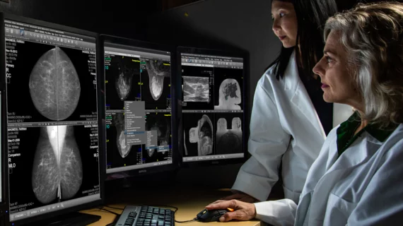
3D mammography volumes have risen in recent years, ballooning read times and leading to physician fatigue and burnout, experts write in JACR. AI presents a potential solution, though real world studies in the U.S. have been small and often lack generalizability, with a need for further evaluation in clinical settings.
Scientists recently aimed to address this knowledge gap, conducting an investigation across four sites involving over 100,000 breast imaging exams. They found clear benefit, with AI increasing the detection of invasive cancers and those found in dense breast tissue while decreasing average stage at diagnosis.
“Our study adds to the recent published evidence that screening outcomes are improved with the implementation of the concurrent use of AI,” Kathy J. Schilling, MD, a breast radiologist with Baptist Health South Florida, Boca Raton, and co-authors concluded. “Further prospective and multicenter studies should be implemented to evaluate AI breast cancer detection in large and diverse populations,” they added.
The study included a total of nine dedicated breast radiologists averaging more than 22 years of experience. Researchers analyzed their results from over 54,000 breast exams (with 339 true positives) logged between 2018–2020 prior to implementing artificial intelligence. These results were put up against findings from another nearly 49,000 exams (369 with true positives), logged between 2020–2022 and backed by AI. Mammograms were gathered uses a GE HealthCare digital breast tomosynthesis system, while the AI tool came from vendor iCAD. (Dr. Schilling was a board member and consultant to the vendor at the time of the study, and other co-authors also had roles there, too.)
The cancer detection rate per 1,000 patients increased by nearly 22%, rising from 6.23 to 7.57 after implementing AI, the study found. Recall rate, or scenarios where women must come back for further imaging, meanwhile, fell from 6.97% to 6.96% after AI. Radiologists also detected more cancers in dense breasts (45% vs. 37.2%), increasing by 7.8%.
Invasive cancer detection per 1,000 also rose by 26%, from 4.63 to 5.83 after AI, and the lobular cancer rate doubled (0.44 to 0.98). The latter, instances of breast cancer that begin in the milk-producing glands, account for about 10% to 15% of invasive cancer cases. Lobular cancer often presents as architectural distortion that are infiltrative in nature, decreasing their detectability on mammograms and often requiring supplemental imaging.
“AI appears to be more sensitive to detection of [architectural distortion] than radiologists,” Schilling and co-authors wrote. “As with all invasive cancers, if [invasive lobular cancer] is not detected until a later stage, the 5-year survival rates decrease. The concurrent use of AI potentially was able to identify these cases more accurately than radiologist interpretation alone.”
Read more, including potential study limitations, in the Journal of the American College of Radiology.

Marty Stempniak has covered healthcare since 2012, with his byline appearing in the American Hospital Association's member magazine, Modern Healthcare and McKnight's. Prior to that, he wrote about village government and local business for his hometown newspaper in Oak Park, Illinois. He won a Peter Lisagor and Gold EXCEL awards in 2017 for his coverage of the opioid epidemic.