
Explainable AI boosts breast cancer detection on MRI
Experts have developed an artificial intelligence model capable of improving the detection of breast cancer on MRI exams.
Unlike other models created for the same purpose, this latest version was developed on a large, diverse set of data and offers evidence backing its determinations—something most would agree is critical for clinical deployment.
Experts involved in its development are hopeful their take on explainable AI can reduce the problematic false positive rates common in breast MRI.
“AI-assisted MRI could potentially detect cancers that humans wouldn’t find otherwise. Previously developed models were trained on data of which 50% were cancer cases and 50% were normal cases, which is a very unrealistic distribution,” lead investigator Felipe Oviedo, PhD, a senior research analyst at Microsoft’s AI for Good Lab, said in a statement. “Those models haven’t been rigorously evaluated in low-prevalence cancer or screening populations (where 2% of all cases or less are cancer), and they also lack interpretability, both of which are essential for clinical adoption.”
Oviedo and colleagues collaborated with the Department of Radiology at the University of Washington to create a model that could identify anomalies in both low- and high-cancer-prevalence settings. They utilized a dataset consisting of more than 10,000 contrast-enhanced breast MRIs acquired at the university between 2005 and 2022. The exams introduced the model to varying categories of breast density, enabling it to flag abnormalities in some of the more challenging images.
“Unlike traditional binary classification models, our anomaly detection model learned a robust representation of benign cases to better identify abnormal malignancies, even if they are underrepresented in the training data,” Dr. Oviedo said. “Since malignancies can occur in multiple ways and are scarce in similar datasets, the type of anomaly detection model proposed in the study is a promising solution.”
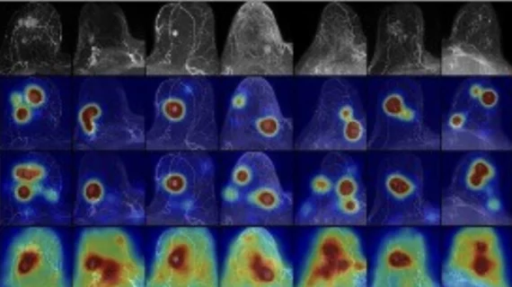
The model outperformed standard benchmark models on both internal and external test sets, and it did so in high and low prevalence tasks as well. It accurately detected abnormalities and provided readers with a heatmap highlighting the area it deemed suspicious. The maps matched biopsy-confirmed lesions, significantly outperforming benchmark models in accurately flagging abnormal exams.
Oviedo and colleagues suggested that, after testing on larger datasets, the model could be used as a triage tool for radiologists with long worklists, enabling them to focus on exams more likely to yield abnormal results.
“Our model provides an understandable, pixel-level explanation of what’s abnormal in a breast,” he said. “These anomaly heatmaps could highlight areas of potential concern, allowing radiologists to focus on those exams that are more likely to be cancer.”
The study abstract is available here.

In addition to her background in journalism, Hannah also has patient-facing experience in clinical settings, having spent more than 12 years working as a registered rad tech. She began covering the medical imaging industry for Innovate Healthcare in 2021.