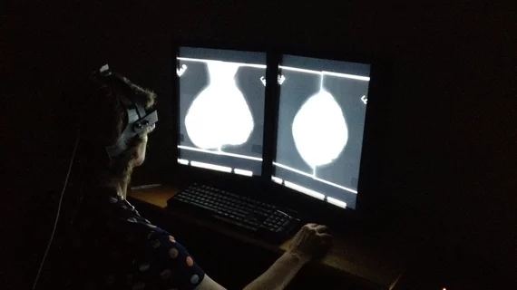
Google AI model ‘impressive’ in detecting breast cancer, besting radiologists
An artificial intelligence system developed by Google is showing “impressive” results in more accurately detecting breast cancer when compared to its human radiologist counterparts.
The tech company has been working alongside Northwestern University and several U.K. institutions for years to develop the model, using thousands of mammography images. Early results—highlighted Jan. 1 in Nature—show that AI spotted breast cancer with greater accuracy and fewer false/negative positives than imaging experts.
“There are some promising signs that the model could potentially increase the accuracy and efficiency of screening programs, as well as reduce wait times and stress for patients,” Shravya Shetty, Google’s technical lead on the project, wrote in a blog post Wednesday.
“But getting there will require continued research, prospective clinical studies and regulatory approval to understand and prove how software systems inspired by this research could improve patient care,” she added later.
Google and its partners trained the AI model using de-identified mammogram images from more than 90,000 women in the U.K. and U.S., then evaluated it on data from 28,000 more women. Specific to the United States study population, the model produced a 5.7% reduction in false positives and a 9.4% reduction in false negatives.
In a separate experiment, scientists trained the model using only data from the United Kingdom, and then tested its skills on the U.S. population. Shetty and colleagues found a 3.5% drop in false positives and 8.1% reduction in false negatives, “showing the model’s potential to generalize to new clinical settings while still performing at a higher level than experts.”
These results are even more noteworthy, Shetty said, as radiologists had access to patients’ history and previous mammograms, while the AI model made its determinations solely based on anonymous images.
In a corresponding Nature opinion piece, American College of Radiology Chief Research Officer Etta Pisano, MD, called the results “impressive,” despite any shortcomings. Those limitations included only incorporating one type of mammography technology and obtaining images from a system developed by a single manufacturer.
“[Google machine learning engineer Scott Mayer] McKinney and colleagues’ results suggest that AI might some day have a role in aiding the early detection of breast cancer, but the authors rightly note that clinical trials will be needed to further assess the utility of this tool in medical practice,” wrote Pisano, who is also a Harvard Medical School professor in residence. “The real world is more complicated and potentially more diverse than the type of controlled research environment reported in this study.”

