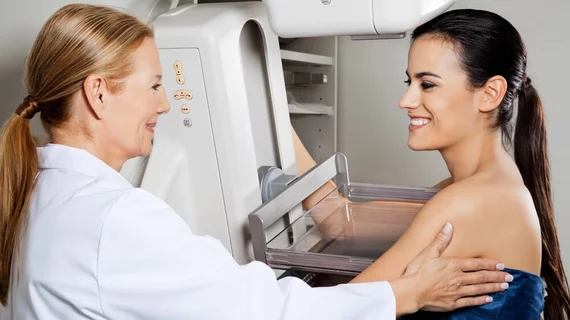
Should women 30-39 years old with focal breast symptoms undergo US, mammography or both?
Targeted breast ultrasound should be the initial imaging evaluation for women between the ages of 30 and 39 presenting with focal breast symptoms, according to a new study published in the American Journal of Roentgenology. Mammography can provide some value in detecting cancers distant from the area of clinical concern, yet its cancer detection rate (CDR) in such a scenario is still rather low.
There are conflicting opinions on how to care for women aged 30-39 presenting with focal breast symptoms. According to the U.K.’s best practice diagnostic guidelines for patients presenting with breast symptoms and the European guidelines for quality assurance in breast cancer screening and diagnosis, ultrasound should be the modality of choice for those women. The U.S. National Comprehensive Cancer Network’s guidelines, on the other hand, recommend mammography.
The authors noted that ultrasound has proven to be quite effective at detecting cancers in women from this age group. But could mammography still provide added value?
“Targeted breast ultrasound is highly sensitive for cancer detection at the site of focal symptoms in this patient population,” wrote Ying Chen, MD, with the Department of Radiological Sciences at the University of Oklahoma Health Science Center in Oklahoma City, and colleagues. “That the negative predictive value approaches 100 percent is an argument for ultrasound as the initial imaging examination in this clinical scenario. Therefore, the primary role and value of bilateral mammography would be to detect cancers distant from the area of clinical concern.”
To test mammography’s value at identifying such cancers, Chen et al. examined data from more than 4,000 diagnostic examinations on more than 3,900 women ages 30-39 years old. All women underwent mammography and ultrasound to evaluate symptoms between June 2006 and August 2016.
Overall, 68 breast cancers were diagnosed in these patients, a CDR of 15.4 per 1,000 exams. Sixty exams led to biopsy of a finding distant from the area of clinical concern, yielding nine incidental malignancies and a CDR of 2 per 1,000 exams. Also, more than 66 percent of the women diagnosed with cancer had “identifiable risk factors,” according to the study.
These findings, the authors conclude, support “recommendations for targeted breast ultrasound as the initial imaging evaluation of women 30–39 years old who have symptoms and support bilateral mammography added to ultrasound for women with risk factors for breast cancer and as an adjunct examination when suspicious or indeterminate sonographic findings are present.”

