In a study of more than 250 COVID-positive patients with a history of any cancer, fewer than half the cohort had chest CT findings deemed typical for COVID-related pneumonia based on an RSNA classification guide.
Based on this and related results, the team behind the research is advising radiologists to consider a COVID diagnosis even when chest CT findings seem to suggest otherwise—as with findings deemed indeterminate, atypical or negative—in patients with preexisting cancers of various kinds.
The work was conducted at Tehran University of Medical Sciences in Iran and published Feb. 6 in BMC Medical Imaging [1].
Retrospectively reviewing the records of 266 patients who met the study criteria, senior study author Kazem Zendehdel, MD, PhD, and colleagues found atypical or indeterminate chest CT findings in 43% of cases.
Some 14% had normal initial chest CT.
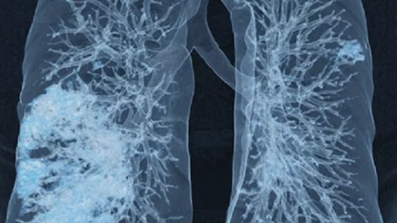
The most commonly reported findings were consolidation mixed with ground-glass opacity (44%), pleural effusion (34%) and pure ground-glass opacity (19.5%).
The most frequently appearing types of cancer were gastrointestinal (29%), hematologic (26%) and breast (10.5%).
Further, risk of death was higher among those who had typical COVID findings on chest CT and those who had a severity score higher than 18, while poorer prognoses were associated with the presence of consolidation, pleural effusion, centrilobular nodules and architectural distortion.
In their discussion, Zendehdel and co-authors underscore that indeterminate results were significantly higher in breast cancer in comparison with other cancers.
In addition, pattern analysis of chest CT features in different cancers revealed that metastatic nodules are “sizably more common in breast cancer,” although pleural effusion was higher in lung cancer.
“These findings highlight the significance of correlating chest CT results with previous imaging, clinical and epidemiological data, and polymerase chain reaction (PCR) test results to make the correct diagnosis,” Zendehdel et al. write.
Among the study’s limitations, the authors acknowledge a dearth of data to compare cancer patients’ active-COVID and pre-COVID CT scans.
The authors state they lacked access to prior chest imaging of most patients who were admitted with COVID. They encourage future researchers to explore this aspect.
The study is posted in full for free.

Dave P. has worked in journalism, marketing and public relations for more than 30 years, frequently concentrating on hospitals, healthcare technology and Catholic communications. He has also specialized in fundraising communications, ghostwriting for CEOs of local, national and global charities, nonprofits and foundations.